

24 September 2021, via Shahriar Lahouti. Last Update 13 June 2026.
CONTENTS
- Preface
- Pathophysiology of stroke
- Physiology: Core infarct vs. ischemic Penumbra
- Clinical presentation
- Anatomy of stroke
- Differential diagnosis
- General approach to diagnosis and management
- Disabling AIS Management
- General supportive early management
- Reperfusion therapy
- Intravenous thrombolytic
- Agents
- Decision-making: Indications & contraindications
- Consent
- Administration
- Recent Updates & Special Situations
- Thrombolytic complication and management
- Endovascular Thrombectomy (EVT)
- Bridge EVT
- Intravenous thrombolytic
- Other therapeutic measures
- Nondisabling Strokes and TIAs
- Media
- Going further
- References
AIS, acute ischemic stroke
CAA, cerebral amyloid angiopathy
CVA, cerebrovascular accident
CVT, cerebral vein thrombosis
DWI, diffusion-weighted imaging
EVT, endovascular therapy
FLAIR, fluid-attenuated inversion recovery
ICH, intracranial hemorrhage
IVT; intravenous Thrombolytics
NCCT, noncontrast computed tomography
CTA, CT angiography
LSN, Last seen normal
LVO, large vessel occlusion
MRI, magnetic resonance imaging
MRA, MR angiography
MT, mechanical thrombectomy;
Preface
Cerebrovascular disease encompasses a variety of medical conditions that affect the cerebral circulation and brain function. Stroke is generally defined as any disease process that interrupts blood flow to the brain. In the following discussion, acute ischemic stroke (AIS) and transient ischemic attack are reviewed in the light of recent guidelines.
Pathophysiology of stroke
The significance of understanding the pathophysiologic classification (Figure 1) is that each stroke type has a distinct presentation, treatment, and prognosis. Acute cerebrovascular syndrome (ACVS) is a constellation of signs and symptoms that are caused by any acute vascular pathology (arterial or venous) that injures the central nervous system tissues. Arterial CVA is classified broadly into two major types: ischemic (≈85%) and hemorrhagic (≈15%) 1. Ischemic stroke (the main focus of this discussion) has three main subtypes: thrombotic, embolic, and hypoperfusion.
Note: Venous CVA (CVT) represents <1% of all strokes and can present with headache, seizure, focal deficits, or altered consciousness. Diagnosis requires MRV/CTV. Anticoagulation is mainstay even with hemorrhagic transformation.
Physiology: Core infarct vs. Penumbra infarct
◾️Core Infarct
- The infarct core refers to tissue that has already been irrevocably damaged. Even if the vessel could be immediately opened, the core infarct would not recover.
- The radiological definition of core infarct is based on the development of cytotoxic edema, reflecting neuronal cell swelling.
- This cytotoxic edema causes diffusion restriction on MRI, the reference standard for defining the core infarct.
- However, CT techniques can also identify the core infarct.
- For anterior hemispheric infarctions, a core infarct volume > ~50-70 ml suggests a poor responsiveness to endovascular therapy (EVT) *.
◾️Ischemic Penumbra
- Ischemic penumbra is tissue that surrounds the core infarct. The ischemic penumbra is malperfused and nonfunctional, yet the tissue remains potentially viable if the blood supply can be restored. The ischemic penumbra is often maintained by a trickle of blood flow supplied via collateral circulation.
- The entire purpose of revascularization (either with thrombolysis or endovascular therapy) is to resurrect the ischemic penumbra.
- Alternatively, if the ischemic penumbra is small, then there is little more tissue to salvage: the stroke has already completed *.
◾️Pace of Progression: Rapid progressor vs. slow progressor
- The natural history of untreated stroke is for the ischemic penumbra to gradually necrose. Thus, over time, the ischemic penumbra will disappear while the core infarct expands.
- The velocity with which the ischemic penumbra necroses varies widely between patients, depending on collateral circulation *.
- Rapid progressors may rapidly complete infarction within hours due to poor collateral flow.
- Slow progressors may continue to have large ischemic penumbras for many hours (or even days). These slow progressors may remain good candidates for revascularization therapy well beyond traditional thrombolysis time windows (e.g., >> 4.5 hours).
- The mantra of stroke neurology is that “time is brain.” However, this is only partially true because different patients progress at different speeds over time. Thus, 4.5 hours could have an entirely different meaning for a slow progressor versus a rapid progressor.
- Applying a rigid time threshold across all patients made sense in the 1990s, when that was all we had. However, in the modern era of neuroimaging, it is increasingly possible to rapidly determine the amount of salvageable tissue.
- Given the ongoing evolution of CT and MRI technology, tissue viability may largely replace time cutoffs when assessing candidacy for interventions.
Clinical Presentation
Acute ischemic stroke (AIS) and transient ischemic attack (TIA) are medical emergencies that occur as a result of a disruption in blood flow to the brain, and originate from a similar pathophysiologic process along the spectrum of acute cerebrovascular syndrome. The core feature of ischemic cerebrovascular accidents is the abrupt onset of focal neurologic deficit (FND). However, the reverse is not true: not all neurological signs and symptoms with sudden-onset FND are ischemic (see DDx below).
◾️Nature of symptoms
- The symptoms and signs in ischemic attack are ‘negative’ in nature (i.e., loss of normal brain function), hence called “Neurologic Deficit”; as opposed to positive or irritative phenomena, which usually have a non-vascular cause (e.g, migraine, seizure, radiculopathy, etc).
- Negative symptoms may include loss of vision, hearing, feeling, or the ability to move a part of the body.
- Positive symptoms suggest active discharge from the central nervous system, including visual (eg, bright lines, shapes, objects), auditory (eg, tinnitus), somatosensory (eg, paresthesia), or motor (eg, jerking movements).
◾️Focal symptoms
- Denotes dysfunction at an anatomically localized brain area. In the context of ischemic events, the focal signs and symptoms are negative in nature; they are called ‘FND’ such as unilateral motor or sensory deficit.
- For example, a focal ischemic insult in the frontal lobe cannot cause bilateral motor deficit or any sensory deficit.
- Examples of non-focal deficits may include altered level of consciousness, bilateral motor or sensory deficits, or amnesia.
- Since focal neurologic deficit can be caused by different pathophysiologic processes (ischemic stroke, hemorrhage, local effect of a mass lesion such as a tumor or an enlarging cerebral artery aneurysm, herniation syndromes, etc.), it is more helpful to consider ‘FND’ in correlation with a vascular territory.
◾️Onset
- Thrombotic stroke has a gradual onset, while embolic stroke and SAH have an abrupt onset.
- Most patients with ‘ICH’ have a gradual onset (sometimes they may have an abrupt onset with maximal deficit severity at the very beginning).
- The negative symptoms in ischemic vascular accidents may involve one or more modalities or functions (e.g., motor +/- sensory).
- The key feature is that it all happens simultaneously at the very onset of attack (while, for example, in migraine aura, the symptoms typically progress from one modality to another; i.e., after the visual symptoms clear, paresthesia begins (when paresthesia is clear, aphasia or other cortical function abnormalities may develop).
◾️Course & Progression
- The neurologic deficits in thrombotic stroke have stuttering (waxing and waning) patterns.
- ‘ICH’ has progressive patterns and severity of signs and symptoms that progress over time.
- Deficits in patients with embolic stroke may suddenly resolve (since endogenous plasminogen activator may lyse the clot).
◾️Duration
- The duration of the neurologic deficit has traditionally been used to separate ‘TIA’ from infarction.
- Typically, the signs and symptoms of TIA last less than 1-2 hours (by classic ‘time-based’ definition < 24 hours). However, with the advent of neuroimaging, it has been shown that time is not reliable to distinguish ‘TIA’ from stroke, as 33% of patients whose neurologic symptoms have lasted for < 24 hours and have been resolved by the time of evaluation, have shown evidence of brain tissue infarction in diffusion-weighted MRI. These patients are at a higher risk of developing a full-blown ischemic infarction.
- In the ‘tissue-based’ definition, TIA is a transient episode of neurologic dysfunction caused by focal ischemia of the brain, spinal cord, or retina, without acute infarction 3. The clinical significance of the ‘tissue-based” definition relies on two facts:
- All patients suspected of having acute cerebrovascular syndrome should have early neuroimaging
- Patients with acute cerebrovascular syndrome (brief and spontaneously resolving) whose neuroimaging shows evidence of infarction have a higher risk of developing ischemic CVA within the next 48hours.
◾️Associated Symptoms
Certain associated symptoms can suggest specific stroke subtypes:
- Altered level of consciousness
- Despite the broad differential diagnosis of altered level of consciousness, when other clinical presentations are suggestive of stroke, reduced alertness can be caused by the following process:
- Elevated ICP secondary to ICH, SAH, CVT, massive embolic or thrombotic stroke (a large hemispheric infarct) is typically followed by edema that can progress to coma.
- Posterior circulation large arteries thrombotic or embolic stroke (In particular, ischemia involving the tegmentum of the pons can cause loss of consciousness)
- Thalamic or pontine hemorrhage
- 📍Patients with signs and symptoms of systemic hypoperfusion (i.e., shock state) can present with an altered level of consciousness, pallor, sweating, tachycardia or severe bradycardia, and low blood pressure. The neurologic signs are typically bilateral, although they may be
- Despite the broad differential diagnosis of altered level of consciousness, when other clinical presentations are suggestive of stroke, reduced alertness can be caused by the following process:
- Seizure
- The occurrence of a seizure within the acute phase of stroke suggests a cortical lesion.
- It is most commonly seen with SAH, ICH (lobar ICH), CVT, and, rarely, may be seen with ischemic stroke (brain embolism).
- Headache
- The presence of headache is often more consistent with other diagnoses (ICH, SAH, CVT) rather than ischemic stroke (rarely, some patients may have headaches in the prodromal period before thrombotic strokes).
- Vomiting
- It is more suggestive of elevated intracranial pressure (ICH, SAH, CVT) or may be seen in patients with posterior circulation large artery ischemia.
- Fever
- Raise the possibility of endocarditis and embolic stroke.
- Chest pain
- Patients with aortic dissection (type A) may develop acute ischemic stroke, which is preceded by the sudden onset of chest pain.
- Rarely, patients with extensive atherosclerosis may develop ‘AIS’ and acute myocardial infarction simultaneously!
- Neck pain
- It may suggest cervicocerebral arterial dissection.
- Elevated ICP
- Global symptoms of elevated ICP include headache, depressed global consciousness, and vomiting.
- Focal symptoms of elevated ICP may be caused by local effects in patients with mass lesions or by herniation syndromes (e.g., subfalcine, central transtentorial, uncal transtentorial, upward cerebellar, cerebellar tonsillar/foramen magnum, and transcalvarial).
- Signs of elevated ICP include CN VI palsies and papilledema.
- Nowadays, bedside ultrasound is widely available, and elevated ICP can be easily identified. Ocular sonography can provide a noninvasive measure of optic nerve sheath diameter, which has been found to correlate with ICP. Several studies have found that diameters of 5 to 6 mm can discriminate between normal and elevated ICP in patients with intracranial hemorrhage and traumatic brain injury (more on this here).
- The etiologies of elevated ICP are discussed here. For this discussion, the presence of intracranial hypertension may suggest hemorrhagic infarction or a massive ischemic infarct.
◾️Other distinguishing features of ischemic stroke
- One may distinguish ischemic vascular accidents from other disease processes by investigating certain historical and clinical features (the table below).
| Stroke type | Major causes | Clinical considerations |
|---|---|---|
| Ischemic | ||
|
Thrombotic Large cerebral arteries – Atherosclerosis – Dissection, such as cervical artery dissection (carotid and vertebral) – Arteritis/vasculitis – Fibromuscular dysplasia, Takayasu arteritis, Giant cell arteritis (large extracranial vessels) Small cerebral arteries (penetrating arteries)
– Arteriolosclerosis – Branched atheromatous disease Blood and coagulation disorders (associates with both venous, e.g., CVT, and arterial thrombosis)
– Sickle cell disease – Polycythemia vera – Essential thrombocytosis – Antiphospholipid syndrome – HIT – Other hypercoagulable state: Protein C or S deficiency, Prothrombin gene mutation, Factor V Leiden, Antithrombin III deficiency, Hyperhomocysteinemia |
– Risk factors for atherosclerosis present – May have history of TIA – Onset of symptoms: gradual – Progression of symptoms: Stuttering (wax and wane) – Lacunes develop over hours or at most a few days – Large artery ischemia may evolve over longer periods For CVT: Headache (most common), seizures, focal deficits (often bilateral or fluctuating), altered consciousness. Diagnosis requires MRV/CTV. Anticoagulation is mainstay even with hemorrhagic transformation. |
|
|
Embolic Cardiac sources with high primary risk for ischemic stroke – Atrial fibrillation, atrial flutter – Sick sinus syndrome – LA thrombus, LV thrombus – Bioprosthetic and mechanical heart valves – Mitral stenosis or rheumatic valve disease – Endocarditis (infective or non-bacterial thrombotic) – Recent MI (within one month prior to stroke) – Chronic MI (together with low EF <28%) – DCM (with LV EF <40%) Cardioartic sources with low or uncertain primary risk for ischemic stroke
Cardiac sources of embolism – Mitral annular calcification – Patent foramen ovale (PFO) – Atrial septal aneurysm w/o PFO – LV aneurysm without thrombus – Left atrial spontaneous echo contrast (“smoke”) – Congestive heart failure with ejection fraction <30% – Apical akinesia or wall motion abnormalities (hypokinesia, akinesia, dyskinesia) other than apical akinesia – HCM or LV hypertrophy – LV hypertrabeculation/non-compaction Aortic source of embolism
Complex atheroma in the ascending aorta or proximal arch (protruding with >4 mm thickness, or mobile debris, or plaque ulceration) Non-cardioartic sources
Fat embolism, Septic embolism |
– Risk factors for atherosclerosis present – History of heart disease – Precipitation: can be precipitated by getting up at night to urinate or sudden coughing or sneezing – Onset of symptoms: Sudden – Progression of symptoms: Deficit is maximal at onset. clinical findings may improve quickly – Unlike thrombotic strokes which often involve a single vascular territory; in embolic stroke multiple sites within different vascular territories may be affected when the source is the heart or aorta. |
|
|
Hyperperfusion – Cardiac arrest – Shock state: Cardiogenic, Hypovolemic, Obstructive, Distributive |
– Symptoms may wax and wane with hemodynamic factors – Diffuse injury pattern in watershed regions |
|
| Hemorrhagic | ||
|
ICH Primary ICH Lobar ICH – Cerebral amyloid angiopathy (CAA) – Cerebral vascular malformations Deep ICH – Hypertensive vasculopathy – Cerebral vascular malformation Secondary ICH (could be either lobar or deep)
– Ischemic CVA hemorrhage transformation – CVT hemorrhage transformation – Brain tumor hemorrhage – Iatrogenic anticoagulation, thrombolytics – Drugs: Cocaine, amphetamine use – Bleeding disorder, liver disease – Posterior reversible encephalopathy syndrome |
– Risk factors: Advanced age, smoking, HTN, bleeding diatheses, illicit drugs, vascular malformations – Precipitation: May be precipitated by sex or other physical activity – Onset: Often gradual but sometimes abrupt – Progression: Signs and symptoms progress during minutes or hours in most patients, but sometimes is maximal at onset – Patients may have reduced alertness |
|
|
SAH (Non-traumatic) – Aneurysmal SAH – Non-aneurysmal SAH (NASAH) – Perimesencephalic nonaneurysmal subarachnoid hemorrhage – Vascular malformation rupture – Other disease that can cause SAH: Intracranial arterial dissection, CVT, Pituitary apoplexy, Bleeding disorders and anticoagulant therapy, cocaine abuse, CAA |
– Risk factors: Smoking, HTN, genetic susceptibility (eg, polycystic kidney disease, family history of SAH) and sympathomimetic drugs (eg, cocaine) – Precipitation: May be precipitated by sex or other physical activity – Onset: Abrupt onset of severe headache – Patients may have reduced alertness – FND are less common than with other types of stroke |
|
Abbreviations & explanations:
HIT: heparin induced thrombocytopenia; HTN: hypertension; LV: left ventricle; LA: left atrium; EF: ejection fraction; DCM: dilated cardiomyopathy; HCM: hypertrophic cardiomyopathy; LVH: left ventricular hypertrophy; CVT: cerebral vein thrombosis.
Factor V Leiden: inherited thrombophilia (activated protein C resistance) associated with increased risk of venous thrombosis, including cerebral venous thrombosis and, less commonly, arterial ischemic stroke.
Lobar ICH: intracerebral hemorrhage located in the cerebral lobes (frontal, parietal, temporal, occipital), typically associated with cerebral amyloid angiopathy (CAA) or vascular malformations; distinct from deep (basal ganglia, thalamus, pons, cerebellum) ICH usually related to hypertensive vasculopathy.
Anatomy of stroke
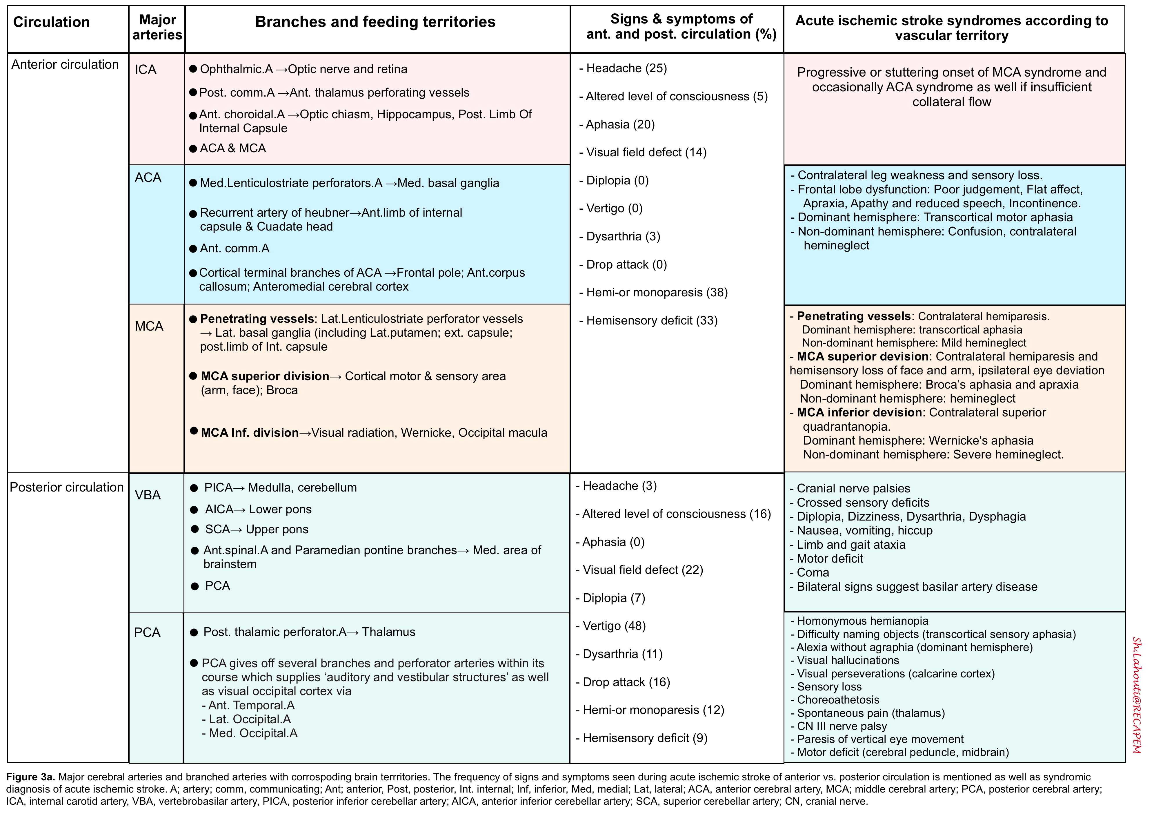
The negative symptoms of ischemic stroke should usually correlate with a specific vascular territory. The cornerstone of diagnosis and appropriate care of patients with acute stroke is to recognize the location and extent of the lesion.
The brain neuroanatomy and relevant vascular supply are shown below (see following slides).
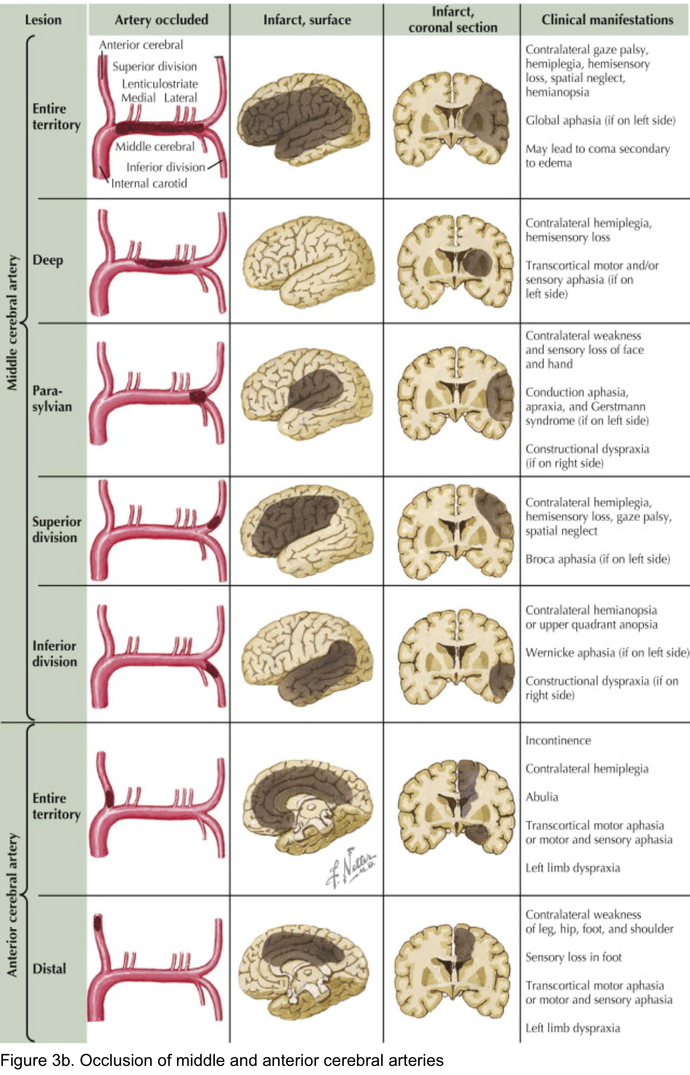
Arterial territory
The arterial territories of the anterior circulation (ACA, MCA) and the posterior circulation (PCA, vertebrobasilar artery) are shown below (Figure 2).
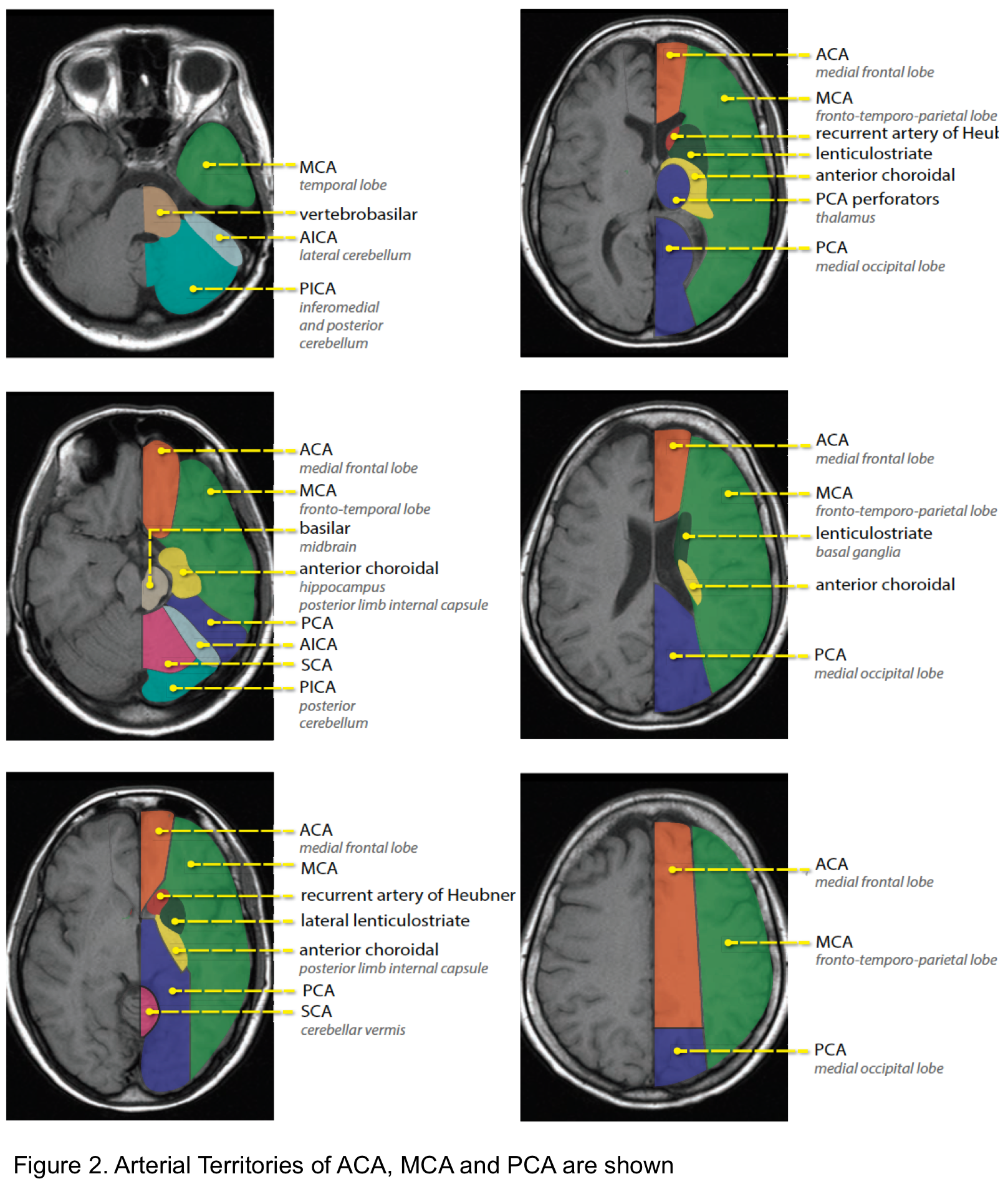
Territorial signs and symptoms
- Findings of acute ischemic stroke in the anterior and posterior circulation are summarized in the following table (Figure 3a). Schematic presentation of occlusion in ACA and MCA is shown in Figure 3b.
- Note that the degree of collateral circulation may cause variations in the specific clinical symptoms and their severity.
Vessel Diameter-Based Classification in AIS
Stroke classification by involved vessel diameter has clinical implications for reperfusion therapy *.
| Vessel Category | Diameter Range | Involved Vessels | EVT Recommendation |
|---|---|---|---|
| Large Vessel Occlusion (LVO) EVT Indicated |
>2.0 mm Typical adult diameter |
• Internal carotid artery (ICA) terminus • M1 segment of middle cerebral artery (MCA) • Basilar artery (BA) • Vertebral artery (V4 segment, selected) |
Class 1, LOE A Recommended |
| Medium Vessel Occlusion (MeVO) Uncertain / Selected |
1.5 – 2.0 mm | • Dominant M2 segment (supplies ≥50% of MCA territory) • Proximal M2, favorable anatomy |
Class 2a, LOE B-NR Reasonable, benefits uncertain |
| Medium Vessel Occlusion (other) Not Recommended |
0.75 – 2.0 mm | • Nondominant / codominant M2 • M3 (distal MCA branches) • A1, A2 (anterior cerebral artery) • P1, P2 (posterior cerebral artery) |
Class 3: No Benefit, LOE A Not recommended |
| Distal Vessel Occlusion Not Recommended |
0.5 – 0.75 mm | • M4 (cortical MCA branches) • A3, A4 (distal ACA) • P3, P4 (distal PCA) |
Class 3: No Benefit EVT not indicated |
| Lacunar Stroke Not Amenable to EVT |
<0.5 mm 100–400 microns typical |
• Lenticulostriate arteries (basal ganglia, internal capsule) • Thalamoperforators (thalamus) • Pontine paramedian branches (pons) • Recurrent artery of Heubner (caudate) | EVT NOT indicated Medical management only |
🧠 Lacunar Stroke – Key Points:
• Vessel diameter: <0.5 mm (penetrating arteries) – NOT classified as LVO, MeVO, or distal occlusion.
• Mechanism: Small vessel disease (microatherosclerosis, lipohyalinosis, branch atheromatous disease).
• Infarct size: Typically <15 mm (often <10 mm).
• Classic syndromes: Pure motor hemiparesis, pure sensory stroke, ataxic hemiparesis, dysarthria-clumsy hand.
• EVT: NOT indicated – no device can safely navigate vessels this small.
• IVT: May be considered if disabling deficits present within 4.5 hours. For non-disabling lacunar strokes → DAPT preferred (Class 3, No Benefit).
• Secondary prevention: Antiplatelet therapy (aspirin, DAPT if indicated), intensive blood pressure control, statin.
📏 Why Vessel Diameter Matters for Treatment:
• Device compatibility: Thrombectomy devices (stent retrievers, aspiration catheters) are designed for vessels >1.5–2.0 mm. Smaller vessels risk perforation, dissection, and subarachnoid hemorrhage.
• Evidence base: All positive EVT trials (Class 1) enrolled LVO (ICA, M1, basilar). Recent MeVO trials showed no benefit for non-dominant M2 and distal occlusions.
• Lacunar strokes are fundamentally different: they arise from small vessel disease, not large emboli, and are not amenable to endovascular approaches.
✅ Abbreviations: LVO = large vessel occlusion; MeVO = medium vessel occlusion; EVT = endovascular thrombectomy; IVT = intravenous thrombolysis; ICA = internal carotid artery; MCA = middle cerebral artery; ACA = anterior cerebral artery; PCA = posterior cerebral artery; mRS = modified Rankin Scale; DAPT = dual antiplatelet therapy.
⚠️ Clinical takeaway: EVT is recommended only for LVO (ICA terminus, M1, basilar artery). For lacunar strokes and most medium/distal occlusions, EVT is not indicated; medical management with antiplatelets and risk factor control is standard.
Large vessel occlusion (LVO)
◾️LVO stroke (“cortical stroke”) is invariably defined as a severe stroke associated with blockages of the proximal intracranial anterior or posterior circulation *.
- It is the most severe form of acute ischemic stroke, accounting for ~10% of acute ischemic strokes, and is associated with poor outcomes if not treated.
- Inclusive definition of LVO 45
- Anterior circulation: Intracranial ICA, M1, A1
- Posterior circulation: Basilar artery, P1
- An NIHSS ≥6 has traditionally been used to predict a cortical stroke; however, there are several caveats here:
- Approximately 5% of large-vessel occlusions presenting within 3 h of last known well will have an NIHSS <4 *.
- NIHS scoring is complicated and time-consuming in the ED.
◾️Prediction of LVO
- 🩺 Clinical: Look for dramatic arm weakness plus cortical signs (aphasia/dysphasia, neglect, gaze deviation, hemianopia). This constellation implies high LVO probability → CTA ASAP to determine eligibility for EVT and rapid transfer if needed.
- 🧮 Tools: There are multiple clinical LVO tools (FAST-ED, RACE) that are reasonably sensitive (~80–90%) for pre-imaging triage, but lack specificity; when a disabling cortical syndrome is present, pursue vascular imaging even if a scale is “negative”.
- VAN tool (a mnemonic for Vision, Aphasia, and Neglect): A more recent tool, the VAN tool, has been shown to have 100% sensitivity, 90% specificity, 74% PPV, and 100% NPV for large-vessel stroke 46,47. Patient must have weakness plus one or all of the V, A, or N to be VAN positive.
- Step 1- Weakness: ask the patient to raise both arms up for 10 seconds and assess for drift, weakness, or paralysis; if any of these are present, proceed to step 2.
- Step 2- V or A or N
- Visual disturbance: Field cut, diplopia, or blindness
- Aphasia: Expressive (repeat and name 2 objects) or receptive (unable to follow commands, close eyes, or make a fist)
- Neglect: Inability to track to one side, ignoring one side, unable to feel both sides at the same time, or unable to identify own arm
- VAN tool (a mnemonic for Vision, Aphasia, and Neglect): A more recent tool, the VAN tool, has been shown to have 100% sensitivity, 90% specificity, 74% PPV, and 100% NPV for large-vessel stroke 46,47. Patient must have weakness plus one or all of the V, A, or N to be VAN positive.
- CTA is the preferred method for the assessment of “vessel status” in patients with acute cerebrovascular syndrome. Advances in the endovascular treatment of LVO stroke reinforce the need to determine the “vessel status” of suspected stroke patients early *.
LVO syndromes
◾️Anterior cerebral artery syndrome
- Either side
- Contralateral leg weakness and sensory loss.
- Frontal lobe dysfunction (poor judgement, flat affect, apraxia, abulia = apathy, and reduced speech, incontinence).
- Alien hand sign: one hand acts involuntarily
- Non-dominant hemisphere: Acute confusion and contralateral hemineglect.
- Dominant hemisphere: Transcortical motor aphasia (due to involvement of the supplementary motor area). This is similar to Broca’s aphasia, but with preservation of repetition.
◾️Proximal MCA syndromes (occlusion near the base of M1)
- Either side:
- Contralateral hemiplegia involving the face and arm > leg.
- Contralateral hemisensory loss.
- Contralateral hemianopia.
- Ipsilateral conjugate eye deviation.
- Nondominant hemisphere: Hemispatial neglect (generally of the left side) and anosognosia.
- Dominant hemisphere: Global aphasia.
◾️Posterior cerebral artery syndromes
- Either side:
- Hemianopia or superior quadrantanopia.
- Thalamic involvement may cause contralateral sensory loss (ventroposterolateral nuclei) or reduced arousal.
- Uncommonly, it may cause contralateral hemiplegia (due to involvement of the internal capsule).
- Dominant hemisphere:
- Alexia without agraphia (patients can write but not read). This results from infarction of the splenium of the corpus callosum, thereby cutting off visual information from the language processing centers.
- Difficulty naming objects (transcortical sensory aphasia).
- Visual agnosia (inability to describe what an object is used for).
- Altered memory (anterograde amnesia) if the medial temporal lobes are involved.
- Nondominant hemisphere:
- Prosopagnosia (inability to recognize faces).
- Bilateral infarctions: Cortical blindness (Anton syndrome).
◾️Proximal basilar artery syndrome (base of the basilar artery)
- Altered level of consciousness (ranging from somnolence to coma).
- Quadriplegia. May also have “crossed” paralysis (e.g., left face and right limbs).
- Oculomotor abnormalities, which may include horizontal gaze palsy (bilateral or unilateral), internuclear ophthalmoplegia (unilateral or bilateral), one and a half syndrome, skew deviation, gaze paretic nystagmus, or bilateral ptosis.
- Pinpoint pupils.
- Bulbar symptoms, which may include: Facial weakness, dysphagia, dysarthria, and palatal myoclonus.
- Pseudobulbar affect.
- Sensory loss to light touch.
◾️Mid basilar artery (Locked-in syndrome)
- Locked-in state
- Quadriplegia and facial paralysis
- Horizontal gaze palsy (vertical gaze remains intact).
- Dysphagia.
- Vertigo.
- Hearing loss can occur.
◾️Top of the basilar syndrome
- This results in ischemia of the midbrain, thalamus, and occipital lobes (but not the pons). The cerebellum may also be involved via the superior cerebellar artery.
- Altered level of consciousness (ranging from somnolence to coma).
- Pupillary abnormalities (may involve afferents to Edinger-Westphal nucleus, CN3 nucleus, or descending sympathetic system). Pupils may be dilated, mid-position, or small.
- Vertical gaze impairment, internuclear ophthalmoplegia, or skew deviation.
- Hemianopsia or complete cortical blindness.
- Amnesia, agitation, and hallucinations (may involve colors and objects).
- Ataxia, tremor, dysarthria (if the cerebellum is involved).
- Homonymous hemianopsia.
Lacunar stroke
Lacunar infarcts are small (2 to 15 mm in diameter) noncortical infarcts caused by occlusion of a single penetrating branch of a large cerebral artery.
◾️Pure motor hemiparesis, Dysarthria-clumsy hand syndrome, Ataxic hemiparesis
- Symptoms:
- Pure motor: Isolated contralateral face/arm/leg weakness. No sensory signs. Dysarthria and dysphagia may be present.
- Dysarthria-clumsy hand: Dysarthria, facial weakness, slight weakness/clumsiness of the contralateral hand.
- Ataxic hemiparesis: Ipsilateral hemi-body weakness and limb ataxia (that is disproportionate to the weakness).
- Localization:
- Corona radiata (small MCA branches).
- Posterior limb of internal capsule (lenticulostriate arteries, anterior choroidal artery, or perforators from the posterior cerebral artery).
- Cerebral peduncle (small proximal posterior cerebral artery branches).
- Anterior pons (basilar perforators).
◾️Pure sensory stroke (thalamic lacunar stroke)
- Symptoms: Unilateral sensory loss of all modalities in the face, arm, and leg without motor deficit.
- Localization: Infarction of the ventral posterior lateral (VPL) and ventral medial nuclei (VPM), supplied by thalamo-perforators from the posterior cerebral artery.
◾️Sensorimotor stroke (thalamocapsular lacunar stroke)
- Symptoms: A combination of thalamic lacune plus pure motor hemiparesis.
- Localization: Posterior limb of the internal capsule plus either thalamic VPL/VPM or thalamic somatosensory radiation. May result from infarction of the thalamoperforator branches of the posterior cerebral artery, or lenticulostriate arteries.
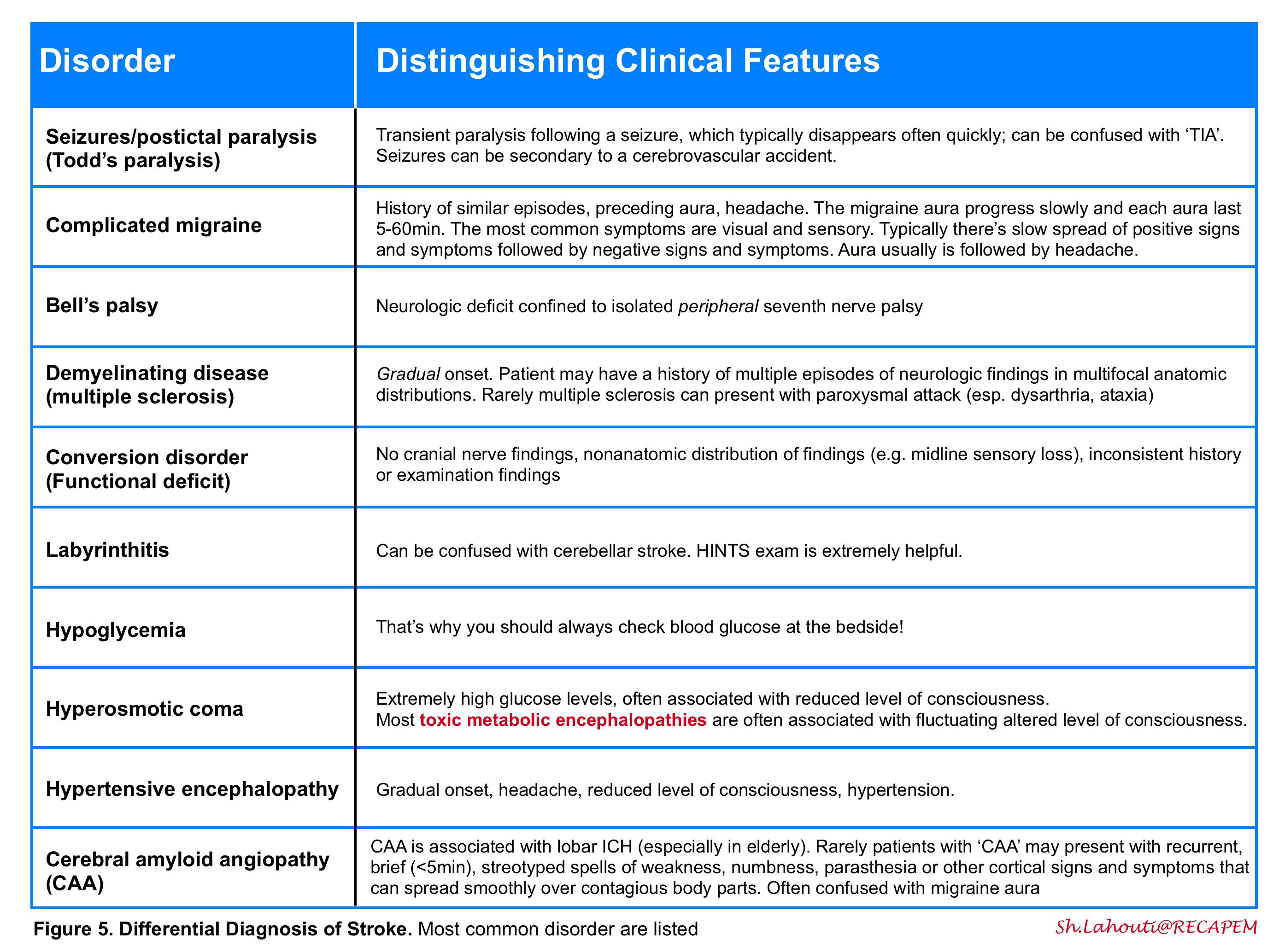
Differential Diagnosis
The history and physical examination should be used to distinguish between other disorders in the differential diagnosis of stroke (Figure 5). As examples, seizures, syncope, migraine, and hypoglycemia can mimic acute ischemia. The most difficult cases involve patients with focal signs and an altered level of consciousness. It is important to ask the patient or a relative whether the patient takes insulin or oral hypoglycemic agents, has a history of a seizure disorder or drug overdose or abuse, medications on admission, recent trauma, or hysteria.
Delineating the duration of attack is helpful to distinguish ischemic accidents from certain other diseases with a paroxysmal nature (e.g., migraine, seizure).
A scoring system (FABS) is proposed for screening and stratifying stroke mimics from acute cerebral ischemia, and to identify patients who may require magnetic resonance imaging to confirm or refute a diagnosis of stroke in the emergency setting 5. This scoring system includes 6 variables with 1 point for each variable, if present:
- Absence of Facial droop
- Negative history of Atrial fibrillation
- Age <50 years
- Systolic Blood pressure <150 mm Hg at presentation
- History of Seizures
- Isolated Sensory symptoms without weakness at presentation
FABS score ≥3 could identify patients with stroke mimics with a sensitivity of 90% and a specificity of 91%. The negative predictive value and positive predictive value were reported as 93% and 87%, respectively.
It seems to be reliable in stratifying stroke mimics from acute cerebral ischemia cases among patients in whom the head CT was negative for any acute findings. It can help clinicians consider advanced imaging for further diagnosis.
Approach To Diagnosis and Management
Overview
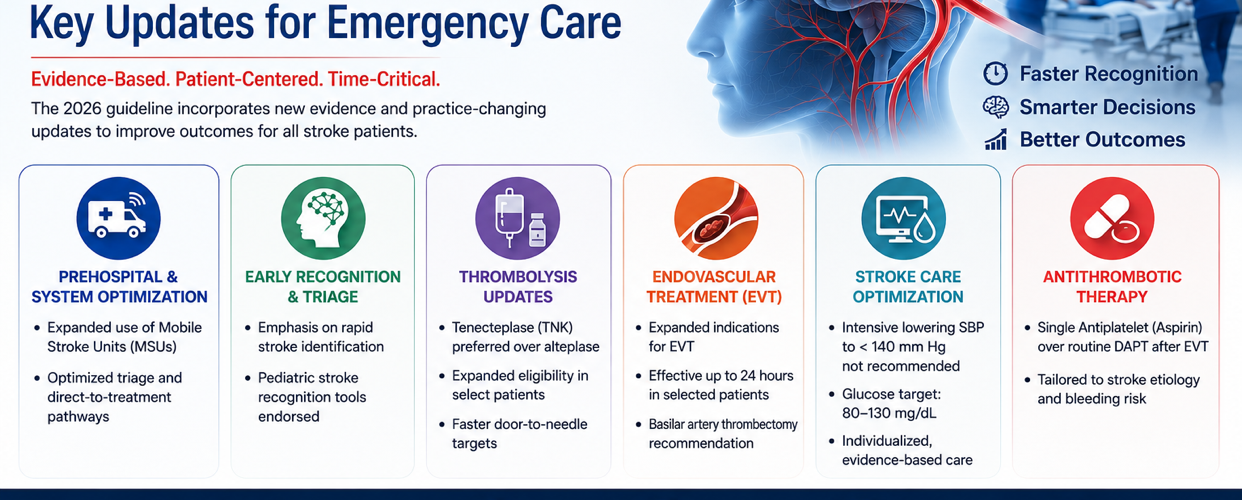
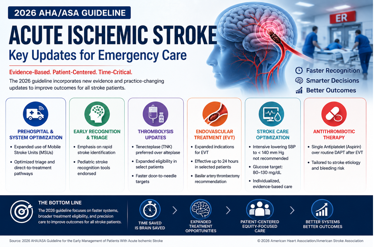
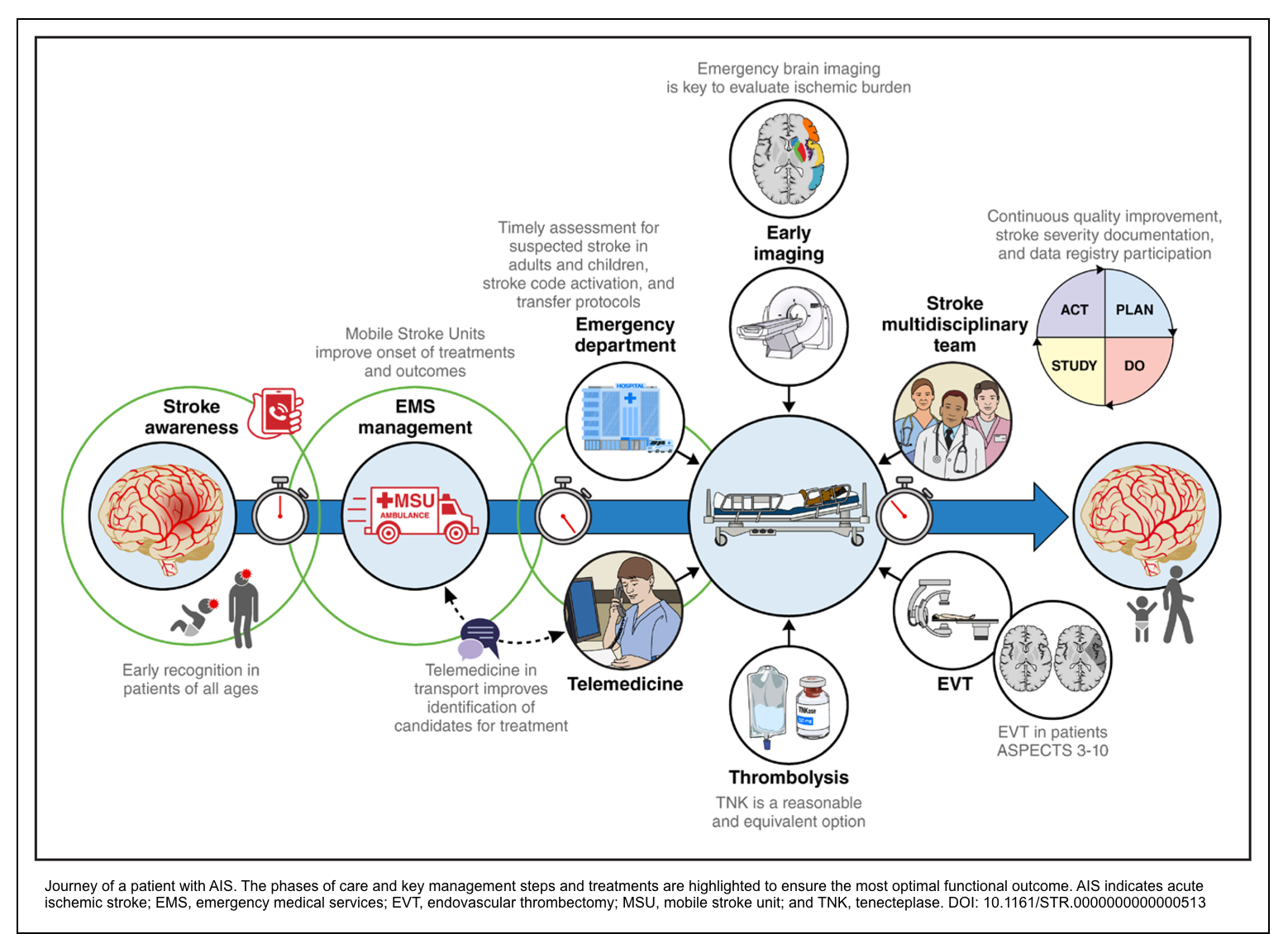
The classic mantra, “time is brain,” explains the current 2026 AHA/ASA stroke guidelines recommendation to enact “an organized protocol for the emergency evaluation of patients with suspected stroke” *.
- The photo (below) walks you through the full journey of a patient with acute ischemic stroke (AIS) — from the moment they call 911, through emergency evaluation and treatment, to in-hospital care *.
- Keep in mind that real-world decisions also depend on local resources, patient preferences, and individual clinical circumstances *.
🚪 Door‑to‑CT/MRI ≤25 min 💉 Door‑to‑needle (IVT) <60 min (target) 🩸 Door‑to‑puncture (EVT) as fast as possible 📋 CT interpretation & review ≤5-10 min 🚑 Scene time ≤15 min (EMS) 🔄 Door‑in‑door‑out (DIDO) for transfer ≤120 min (ideal)
⏱️ Time zero – start clock
• Medical stabilization & history (time last known well)
• Focused exam + NIHSS (disabling vs. non‑disabling)
• Point‑of‑care glucose • Stroke team notification
🎯 Door‑to‑imaging ≤25 min
• Exclude hemorrhage • Assess ASPECTS (for EVT candidacy)
⏱️ CT review & interpretation: target ≤5-10 min
(Unable to perform basic ADLs: walking, dressing, communicating, eating)
• IV thrombolysis recommended (Class 1)
• Do NOT delay for advanced imaging if ≤4.5h
• Either alteplase (0.9 mg/kg) OR tenecteplase (0.25 mg/kg)
• Extended window (4.5‑9h or wake‑up): requires DWI‑FLAIR or CTP mismatch (Class 2a)
• IVT NOT recommended (Class 3, No Benefit)
• Dual antiplatelet therapy (DAPT) preferred
• Urgent workup (MRI/CTA) within 24‑48h
🎯 Door‑to‑needle target: <60 minutes
• Alteplase 0.9 mg/kg (max 90 mg): 10% bolus + 60 min infusion
• Tenecteplase 0.25 mg/kg (max 25 mg): single bolus over 5-10 sec
• Do NOT wait for CTA, CTP, or lab results (unless coagulopathy suspected)
Coverage: aortic arch to vertex (for EVT planning)
Assess for LVO (ICA terminus, M1, basilar), tandem occlusions, collaterals
⏱️ Can be done immediately after NCCT (adds 2-3 min)
(ICA‑T, M1, basilar artery)
• Early window (0‑6h): EVT recommended (Class 1)
• Late window (6‑24h): EVT recommended only with advanced perfusion mismatch (CTP or MR PWI)
• Basilar artery occlusion: EVT up to 24h (NIHSS ≥10, Class 1)
• Large core (ASPECTS 3‑5): EVT recommended (Class 1)
• 🎯 Door‑to‑puncture: as fast as possible (tracked metric)
• Continue medical management
• Antiplatelet therapy (aspirin within 24‑48h)
• DAPT for minor non‑cardioembolic stroke (21 days)
• Investigate etiology (carotid, cardiac, etc.)
• BP target after IVT: maintain SBP <180, DBP <105 (avoid intensive lowering to <140 mmHg – Class 3)
• After successful EVT: avoid SBP <140 mmHg (harmful)
• Glycemia: target 140‑180 mg/dL; intensive control (80‑130 mg/dL) NOT recommended
• Dysphagia screen • DVT prophylaxis (IPC) • Antiplatelet for secondary prevention
• Follow‑up imaging at 24h before starting antiplatelet/anticoagulant
• Target Door‑In‑Door‑Out (DIDO) time: ≤120 minutes (ideal)
• Give IVT if eligible • Obtain CTA if possible • Rapid transfer to thrombectomy‑capable center
• Tenecteplase (TNK) 0.25 mg/kg – equivalent to alteplase (Class 1)
• Disabling vs. non‑disabling – primary driver for IVT (non‑disabling → DAPT)
• EVT for larger core (ASPECTS 3‑5) – Class 1; ASPECTS 0‑2 – Class 2a
• Basilar artery occlusion – EVT up to 24h (NIHSS ≥10, Class 1)
• Extended window IVT (4.5‑9h / wake‑up) – requires DWI‑FLAIR or CTP mismatch (Class 2a)
• Pediatric AIS – IVT may be considered (Class 2b); EVT for age ≥6y (Class 2a)
• BP after reperfusion – intensive SBP lowering to <140 mmHg NOT recommended (Class 3)
• Glycemia – target 140‑180 mg/dL; avoid 80‑130 mg/dL
Time benchmarks summary: Door‑to‑CT ≤25 min | CT interpretation ≤5-10 min | Door‑to‑needle (IVT) <60 min | Door‑to‑puncture (EVT) as fast as possible | DIDO ≤120 min (transfer).
Abbreviations: LVO = large vessel occlusion; ASPECTS = Alberta Stroke Program Early CT Score; CTP = CT perfusion; DWI‑FLAIR = diffusion‑weighted / fluid‑attenuated inversion recovery mismatch; DAPT = dual antiplatelet therapy; MSU = mobile stroke unit (recommended when available); DIDO = door‑in‑door‑out.
History
⏳Timing of onset of symptoms and signs
- A stroke tends to be abrupt
- Ask about and clarify the specific time of onset
- Establish the time of Last Seen Normal” (LSN)
- This anchors the onset time when symptoms are unwitnessed; however, LSN should not be conflated with tissue viability.
- Patients presenting close to 24 h from LSN may still be candidates for late-window EVT if imaging reveals a small infarct core with a substantial penumbra.
- Practically, LSN documents chronology; selection for therapy is driven by imaging-demonstrated salvageable tissue *.
- This anchors the onset time when symptoms are unwitnessed; however, LSN should not be conflated with tissue viability.
- Wake Up” Strokes – Where urgent imaging is paramount
- Definition: A wake-up stroke occurs when the precise onset time is unknown, but the “last seen normal” is when the patient went to sleep.
- Timing of most events: Most wake-up strokes occur just before awakening, driven by physiologic changes (early morning cortisol surge and blood pressure spikes) *.
- Diurnal variations (early morning blood pressure and cortisol surges) suggest that many unwitnessed events occur shortly before awakening, not at bedtime.
- Implication for salvageable tissue: Because the stroke occurs closer to awakening, there is likely more salvageable tissue than if the stroke had occurred immediately upon falling asleep.
- Wake-up strokes have a higher probability of treatment eligibility than witnessed late-window strokes because the true ischemic duration may be short.
- Imaging findings: Advanced imaging (CT perfusion or MRI DWI-FLAIR mismatch) is more likely to reveal salvageable penumbra, making many wake-up stroke patients candidates for reperfusion therapy *.
- 💡Clinical pearl: Unknown onset should prompt, not prevent, reperfusion work-up.
- Do not exclude on time grounds alone: Do not exclude patients from EVT evaluation solely based on uncertain time of onset for wake-up strokes.
- Action item: Instead, expedite advanced imaging for all suspected wake-up strokes to assess for salvageable tissue and determine extended window eligibility.
- Establish the time of Last Seen Normal” (LSN)
- 📍Bottom line
- Wake-up strokes are imaging emergencies.
- For wake-up strokes, unknown onset is NOT a contraindication to EVT. The question is not “When did it start?” but rather “Is there salvageable tissue on imaging?” Expedite CTP or MRI for all suspected wake-up strokes *.
- Use vascular and perfusion/collateral imaging to identify candidates for IV thrombolysis and/or EVT, and keep decisions anchored to disability and salvageable tissue, not the clock *.
- Wake-up strokes are imaging emergencies.
◾️Other components of history
- Risk factors, e.g., DM, HTN, smoking, vascular disease,
- Obtain a history of anticoagulation use and the timing of the last dose taken
- Obtain a history of previous stroke, recent surgery, systemic or intracranial bleeding history, and known intracranial masses
- Think about mimics
- If a patient arrives with the EMS, ask about the pre-hospital screening scale and any change in neurological status since the time of onset.
Physical exam
◾️Assessment of airway, breathing, and circulation is the top priority. Next, the goals of examination are to confirm the diagnosis of stroke, exclude stroke mimics, and identify comorbidities. In cases where time-sensitive treatment decisions must be made, the initial assessment may be very brief and performed in parallel to other interventions.
◾️NIHSS (🧮 MDCalc).
- The National Institutes of Health Stroke Scale is widely used to assess neurological deficits in stroke patients in a structured manner. See media below🎥
- It’s a standardized, 15-item neurological examination that quantifies stroke-related deficits across multiple domains, including level of consciousness, language, neglect, visual fields, motor strength, ataxia, and sensation.
- Each item is scored numerically, with total scores ranging from 0 (normal) to 42 (most severe).
- Higher scores predict a larger lesion size, greater stroke severity, and worsened short- and long-term outcomes.
- All patients should have the ‘NIHSS’ calculated, as it is a widely accepted measure of stroke severity.
- Generally, this should be performed after initial stabilization and neuroimaging *.
- The NIHSS serves 3 primary functions in acute ischemic stroke care:
- It communicates stroke severity among clinicians
- It predicts patient prognosis (higher scores correlate with worse outcomes)
- It helps identify patients with large vessel occlusion who may be candidates for EVT.
📍The Component of a complete neurological examination is discussed here.
- Traditionally, stroke was classified as “minor” (NIHSS ≤5) vs. “major” (NIHSS ≥6), and treatment decisions were based on a numeric cutoff.
- Problem
- NIHSS of 2 from severe aphasia would be disabling, while the same score from facial droop might not be disabling.
- Numeric cutoff fails to capture functional impact *.
- NIHSS of 2 from severe aphasia would be disabling, while the same score from facial droop might not be disabling.
◾️New approach (2026 AHA/ASA Guideline)
- Replace numeric classification (NIHSS) with a functional, patient-centered paradigm based on disabling vs. non-disabling *.
- Defining disabling
- A deficit that prevents a patient from performing basic activities of daily living (bathing, dressing, ambulating, toileting, eating) or returning to work is considered disabling, regardless of the numerical NIHSS score. Conversely, deficits that do not impair these functions are considered non-disabling.
- Classify the presentation as disabling when it threatens independence or meaningful quality of life (speech/language, vision, ambulation, dominant-hand function, or level of consciousness), and nondisabling when deficits are unlikely to impact these domains if untreated *.
- Features typically considered disabling:
- Speech: Major speech deficit (severe aphasia or dysarthria compromising functional communication)
- Motor: Dense motor loss (especially dominant hand/arm, profound leg weakness), or ant weakness limiting sustained effort against gravity.
- Vision: Amaurosis fugax, cortical blindness, dense hemianopia.
- Consciousness/Brainstem: depressed level of consciousness, locked-in syndrome from basilar occlusion *.
- Patient and family context:
- What is disabling varies with occupation, age, and lifestyle.
- Shared decision-making is vital for subtle deficits.
- Implementation
- The practical categorization of “AIS” as disabling vs. nondisabling will reframe the first decision point (Figure below).
- For intravenous thrombolysis (IVT) decisions, the primary driver is no longer the NIHSS score alone *.
- ED teams must weigh the patient’s premorbid status, functional demands, and goals of care.
- The practical categorization of “AIS” as disabling vs. nondisabling will reframe the first decision point (Figure below).
- A deficit that prevents a patient from performing basic activities of daily living (bathing, dressing, ambulating, toileting, eating) or returning to work is considered disabling, regardless of the numerical NIHSS score. Conversely, deficits that do not impair these functions are considered non-disabling.
Inability to communicate
dominant hand / ambulation
blindness, cortical blindness
large hemispheric syndromes
📊The Paradigm Shift
- The following table summarizes the practical change at the first point of decision-making in patients with AIS *.
| Aspect | Traditional Approach (Prior Guidelines) | 2026 Guideline Approach |
|---|---|---|
| Primary driver for IVT decision | NIHSS score (e.g., treat if NIHSS ≥ 6, uncertain if 0-5) | Is the deficit disabling for THIS patient? Functional impact overrides numeric score |
| Role of NIHSS | Decisive — primary gatekeeper for treatment eligibility | Descriptive and prognostic, but not the sole gatekeeper Guides but does not dictate |
| Focus of assessment | Neurological deficits as scored by an examiner | Functional impact on patient’s ability to perform basic ADLs (bathing, dressing, ambulating, toileting, eating) and return to work |
| Problem addressed | A patient with NIHSS = 2 (e.g., severe aphasia or monocular blindness) might be disabled but considered “mild” by score alone → denied treatment | NIHSS guides, but clinical judgment of “disabling” overrides the number → appropriate treatment for disabled patients with low scores |
| Treatment for NIHSS 0-5 | Uncertain, often debated, frequently no treatment given | Clearly defined: • Treat with IVT if deficits are disabling • Do NOT treat with IVT if deficits are non-disabling (use DAPT instead) |
| Treatment for NIHSS ≥6 | IVT recommended | IVT recommended (these are almost always disabling by definition) |
| EVT eligibility | NIHSS ≥ 6 required for most trials | NIHSS ≥ 6 remains standard (disabling almost always implied in LVO patients) |
| Clinical example Two patients, both NIHSS = 2 |
Both would be considered “mild stroke” — treatment decisions inconsistent | Patient A (Disabling – severe aphasia): Give IVT Patient B (Non-disabling – isolated facial droop + mild sensory loss): Do NOT give IVT (DAPT preferred) |
📋 Operational Definition of “Disabling” (2026 Guideline, Table 4):
“If the observed deficits persist, would they still be able to do basic activities of daily living and/or return to work?”
Typically considered DISABLING: Complete hemianopsia, severe aphasia, severe hemi-attention/extinction, any weakness limiting sustained effort against gravity.
Typically considered NON-DISABLING: Isolated mild aphasia (still able to communicate), isolated facial droop, mild hemisensory loss, mild ataxia if patient can still ambulate.
📖 Direct from the 2026 Guideline (Section 4.6.1, Page 38):
• Class 1, LOE A: “In adult patients with AIS with disabling deficits (regardless of NIHSS score), and eligible for IVT, faster treatment improves functional outcomes.”
• Class 3, LOE B-R: “In eligible adult patients with AIS presenting with mild non-disabling stroke deficits (eg, isolated sensory syndrome) within 4.5 hours… IVT is not recommended as it has not shown superiority… compared to double antiplatelet treatment.”
✅ What the NIHSS is STILL used for (not abandoned):
• Communicating stroke severity • Predicting prognosis • Identifying potential LVO (higher scores) • Selecting patients for EVT trials (NIHSS ≥6 remains common) • Measuring neurological change over time
🔑 Key Takeaway: The 2026 guideline introduces a fundamental conceptual shift: For IVT decisions, the primary question is no longer “What is the NIHSS score?” but rather “Is the deficit disabling for this patient?” The NIHSS remains valuable but is no longer the sole gatekeeper for thrombolysis. Patients with low NIHSS scores (0-5) who have disabling deficits should receive IVT, while those with truly non-disabling deficits should not (DAPT preferred).
⚠️ Important: This shift applies primarily to IV thrombolysis. For EVT eligibility, NIHSS ≥6 remains the standard.
Stroke Algorithm
The 2026 AHA/ASA Guideline introduces a fundamental shift in acute stroke triage: the primary decision point is no longer the NIHSS score, but rather whether the neurological deficit is disabling *. The algorithm below provides a practical framework for initial evaluation and disposition:
- Disabling deficits trigger a CODE STROKE activation, regardless of NIHSS score, with STAT NCCT/CTA and immediate consideration for IV thrombolysis or endovascular thrombectomy (EVT), provided the time from last seen normal (LSN) is not clearly >24 hours *.
- Non-disabling deficits require further stratification: patients with persistent deficits or resolved high-risk features (e.g., ABCD² score ≥4, atrial fibrillation, carotid stenosis) need urgent imaging (MRI/CTA) within 24-48 hours, whereas those with low-risk, fully resolved symptoms may be managed with outpatient evaluation *.
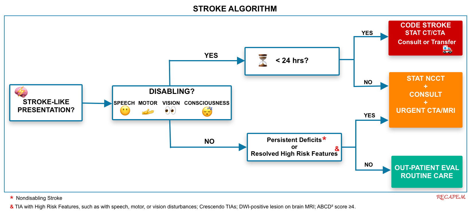
- This is a hospital emergency protocol that mobilizes a dedicated stroke team for rapid evaluation and treatment of suspected acute stroke.
- It is activated for any patient with an acute focal neurological deficit suspicious for stroke, regardless of age, and regardless of whether the time of onset is known or unknown, as long as the time from last seen normal (LSN) is not clearly >24 hours *.
- The goal is door‑to‑imaging ≤25 minutes, door‑to‑needle (IVT) <60 minutes, and rapid EVT assessment. Non‑disabling deficits or low‑risk TIAs usually do not require a full Code Stroke but still need urgent evaluation.
- Do not exclude a patient from Code Stroke activation simply because the onset time is unknown (e.g., wake-up stroke, unwitnessed onset) *.
- Do not refuse to activate Code Stroke just because the patient might be outside the window, because advanced imaging (CTP, MRI) may still show salvageable tissue up to 24 hours (or even 4.5-9 hours for IVT in select patients) *.
- However, once the time from last seen normal (LSN) is clearly known to be >24 hours (e.g., 48 hours), there is no evidence-based reperfusion therapy (IVT or EVT) to offer. Therefore, a full Code Stroke activation is not indicated *.
- Not all acute neurological deficits require STAT NCCT with Code Stroke urgency.
- Only patients with DISABLING deficits who are within potential reperfusion windows (LSN ≤24 hours or unknown) need emergent NCCT (door‑to‑CT ≤25 min) *. Non‑disabling deficits and TIAs require urgent (but not STAT) imaging within 24-48 hours, as they are not candidates for acute reperfusion.
Brain imaging
- All patients suspected of ‘AIS’ and ‘TIA’ (determined to be stable) should be taken immediately for neuroimaging (Non-contrast head CT +/- other modalities) *.
- Brain and neurovascular imaging play a crucial role in acute stroke by 7 8:
- Differentiating ischemia from hemorrhage (Is there an ICH at NCCT that is a contraindication to IVT or EVT, or is there a large well-established hypoattenuating infarct?)
- Excluding stroke mimics, such as a tumor.
- Assessing the status of large cervical and intracranial arteries (is there a proximal LVO seen at CTA that can be treated with EVT?)
- Estimating the volume of brain tissue that is irreversibly infarcted (ie, infarction core)
- Estimating the extent of potentially salvageable brain tissue that is at risk for infarction (ie, ischemic penumbra)
- The approach to imaging may differ according to individual patient characteristics (eg, time from stroke onset, potential candidate for reperfusion therapies) and local availability of stroke expertise and imaging capabilities.
- For example, in patients suspected of ‘TIA’ who are asymptomatic (time is not brain) by the time of presentation to the hospital, it may be reasonable to obtain a brain MRI initially if available.
- However, when a patient is a potential candidate for IVT based on clinical characteristics and initial head CT, do not delay thrombolytic administration while waiting to perform more advanced imaging, such as CTA.
⚠️ A normal early CT or CTA does not exclude ischemic stroke.
- Lacunar infarcts, distal vessel occlusions, and small ischemic cores can be radiographically occult in the hyperacute phase.
- 💡Clinicians should treat the clinical syndrome, and when imaging findings appear discordant with the patient’s presentation, pursue specialist advice rather than prematurely excluding the diagnosis.
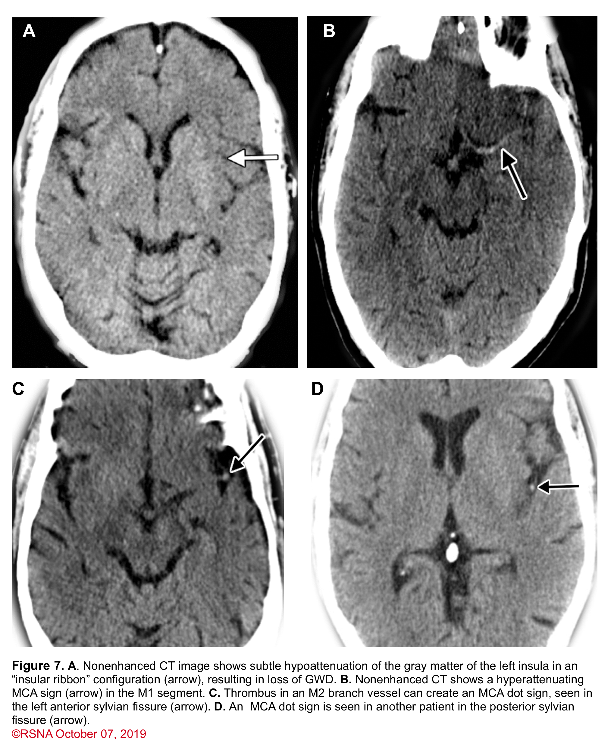
Noncontrast Head CT
- It is the most common imaging modality used for triage of ‘AIS’; since it is widely available, rapid, and can easily detect ‘ICH’.
- Most AIS are not visualized by a non-contrast brain CT in the early hours of a stroke.
- Therefore, the utility of the first brain CT is primarily to exclude ‘ICH’, abscess, tumor, and other stroke mimics, as well as to detect current contraindications to thrombolytics (e.g., extensive regions of clear hypoattenuation)7.
- Minor ischemic changes (i.e., early signs of infarction) on CT are not a contraindication to treatment; these include
- Subtle or small areas of hypodensity
- Loss of gray-white distinction
- Obscuration of the lentiform nucleus
- Presence of a hyperdense artery sign (figure 7).
- In patients with a hyperdense MCA sign, IVT can be beneficial.
- ⚠️IVT is not beneficial in the presence of extensive regions of obvious hypodensity consistent with irreversible injury on initial head CT and is not recommended 6 (Figure 8).
- 📍Severe hypoattenuation is defined as obvious hypodensity, which represents irreversible injury 6. These patients have a poor prognosis despite IVT.

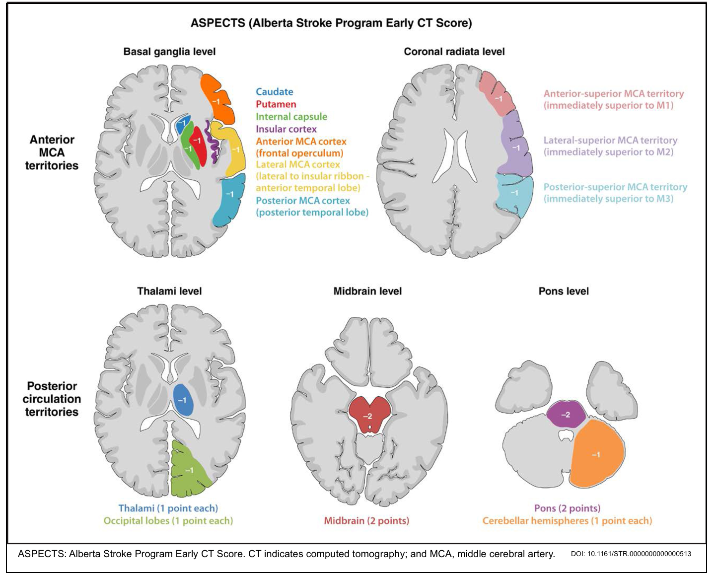
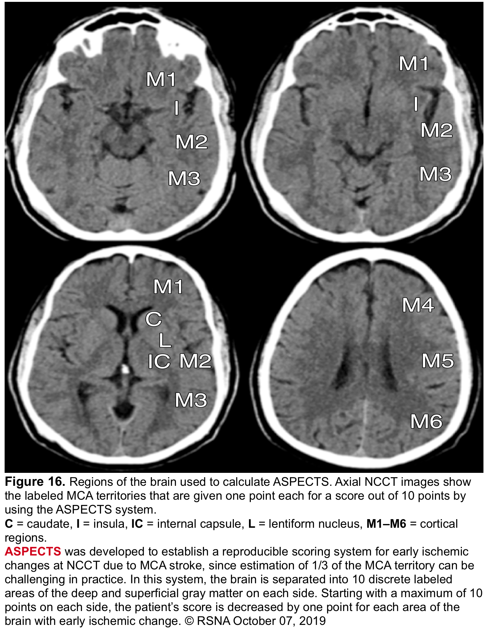
- ASPECTS: Alberta Stroke Program Early CT Score (🧮MDCalc)
- ASPECTS is a standardized 10-point topographic scoring system used to quantify early ischemic changes on non-contrast CT in patients with acute anterior circulation ischemic stroke *.
- It is a simple, reproducible scoring system that assigns points to specific regions of the middle cerebral artery (MCA) territory.
- A normal CT scan receives a score of 10 points.
- One point is deducted for each region showing early ischemic changes (e.g., hypodensity, loss of gray-white matter distinction, sulcal effacement).
- ASPECTS is a standardized 10-point topographic scoring system used to quantify early ischemic changes on non-contrast CT in patients with acute anterior circulation ischemic stroke *.
- Clinical implication
- Identify extensive hypodensity as an absolute contraindication to IVT.
- Select patients for EVT (ASPECTS 3-10 in early window; ≥6 in late window)
- Predict prognosis.
- ASPECTS is simple, fast, and requires only NCCT, but has limitations, including:
- Inter-rater variability.
- Reduced sensitivity in very early strokes (<3 hours)
- For posterior circulation strokes, PC-ASPECTS is used (Below table) *.
- Inability to assess penumbra or collaterals.
- Clinical implications of ASPECT for IVT/EVT
| Clinical Application | ASPECTS / PC-ASPECTS | Recommendation (2026 Guideline) | Notes / Evidence |
|---|---|---|---|
| IV Thrombolysis (IVT) Class 1 / Class 3 |
Any ASPECTS (10 to 0) | • Mild to moderate early ischemic changes (ASPECTS 3-9) are NOT a contraindication – IVT recommended (Class 1, LOE A). • Extensive clear hypodensity (frank hypodensity > contralateral white matter, responsible for clinical symptoms) – IVT NOT recommended (absolute exclusion). |
Section 4.6.1, Recommendation #7 & Table 8 (Page 52). “Clear hypodensity” is when density is less than contralateral white matter. |
| EVT – Early Window (0-6 hours) Class 1, LOE A |
ASPECTS 3 – 10 | EVT is recommended for anterior circulation LVO (ICA or M1) with NIHSS ≥6, prestroke mRS 0-1. | HERMES, MR CLEAN, ESCAPE, SWIFT PRIME, REVASCAT. Advanced imaging (CTP) is optional in early window. |
| EVT – Early Window (0-6 hours) Class 2a, LOE B-R |
ASPECTS 0 – 2 (large core) | EVT is reasonable for selected patients: age <80 years, NIHSS ≥6, prestroke mRS 0-1, no significant mass effect. | LASTE, SELECT2, ANGEL-ASPECT. Functional independence rates lower than in small-core trials (13.3% vs 7.5% with medical therapy). |
| EVT – Late Window (6-24 hours) Class 1, LOE A |
ASPECTS ≥ 6 | EVT is recommended with advanced imaging REQUIRED (CTP or MR DWI/PWI mismatch showing salvageable tissue per DAWN/DEFUSE-3 criteria). | DAWN, DEFUSE-3, AURORA meta-analysis. Requires core <70 mL, mismatch ratio >1.8, mismatch volume >15 mL. |
| EVT – Late Window (6-24 hours) Class 1, LOE A |
ASPECTS 3 – 5 (large core) | EVT is recommended for selected patients: age <80 years, NIHSS ≥6, prestroke mRS 0-1, no significant mass effect, with advanced imaging mismatch. | ANGEL-ASPECT, SELECT2 (extended window large-core trials). Benefit magnitude smaller than in small-core patients. |
| Posterior Circulation (Basilar Artery) Class 1, LOE A |
PC-ASPECTS ≥ 6 | EVT is recommended within 24 hours for basilar artery occlusion with NIHSS ≥10, prestroke mRS 0-1. | ATTENTION, BAOCHE. PC-ASPECTS is a 10-point scale (pons and midbrain = 2 points each; thalami, occipital lobes, cerebellum = 1 point each). For NIHSS 6-9, effectiveness is not well established (Class 2b). |
C = Caudate | L = Lentiform | IC = Internal capsule | I = Insula | M1 = Anterior MCA | M2 = MCA lateral to insula | M3 = Posterior MCA | M4 = Anterior MCA (superior) | M5 = Lateral MCA (superior) | M6 = Posterior MCA (superior)
PC-ASPECTS (Posterior circulation – 10 points): Pons (2), Midbrain (2), Thalami (2), Occipital lobes (2), Cerebellar hemispheres (2).
Scoring: Start with 10, deduct 1 point for each region with early ischemic change (hypodensity, loss of gray-white distinction, sulcal effacement). Lower score = larger infarct core.
• Inter-rater variability (especially for scores 0-2 and 8-10).
• Less sensitive than MRI-DWI for early ischemic changes (<3 hours).
• Not validated for posterior circulation (use PC-ASPECTS instead).
• In late window (6-24h), advanced perfusion imaging (CTP or MR PWI) is REQUIRED – ASPECTS alone is insufficient.
✅ Key takeaway: Mild to moderate early ischemic changes (ASPECTS 3-9) are NOT a contraindication to IVT. EVT is recommended for ASPECTS 3-10 in early window (0-6h) and ASPECTS ≥6 in late window (6-24h) with advanced imaging. Large core (ASPECTS 0-5) patients may still benefit from EVT (Class 1 or 2a). PC-ASPECTS ≥6 is required for basilar artery EVT.
Brain MRI
- Standard brain MRI protocols that include conventional T1-weighted, T2-weighted, FLAIR, DWI, and the apparent diffusion coefficient (ADC) map can reliably diagnose both AIS and acute hemorrhagic stroke in emergency settings.
- Major drawbacks of MRI are that it is not readily available and its use may be limited by contraindications (e.g., metal implant or pacemakers) or patient intolerance (i.e. claustrophobia). Newer ultrafast MRI protocols can reduce acquisition times from the 15 to 20 min required for conventional MRI to 5 min or less.
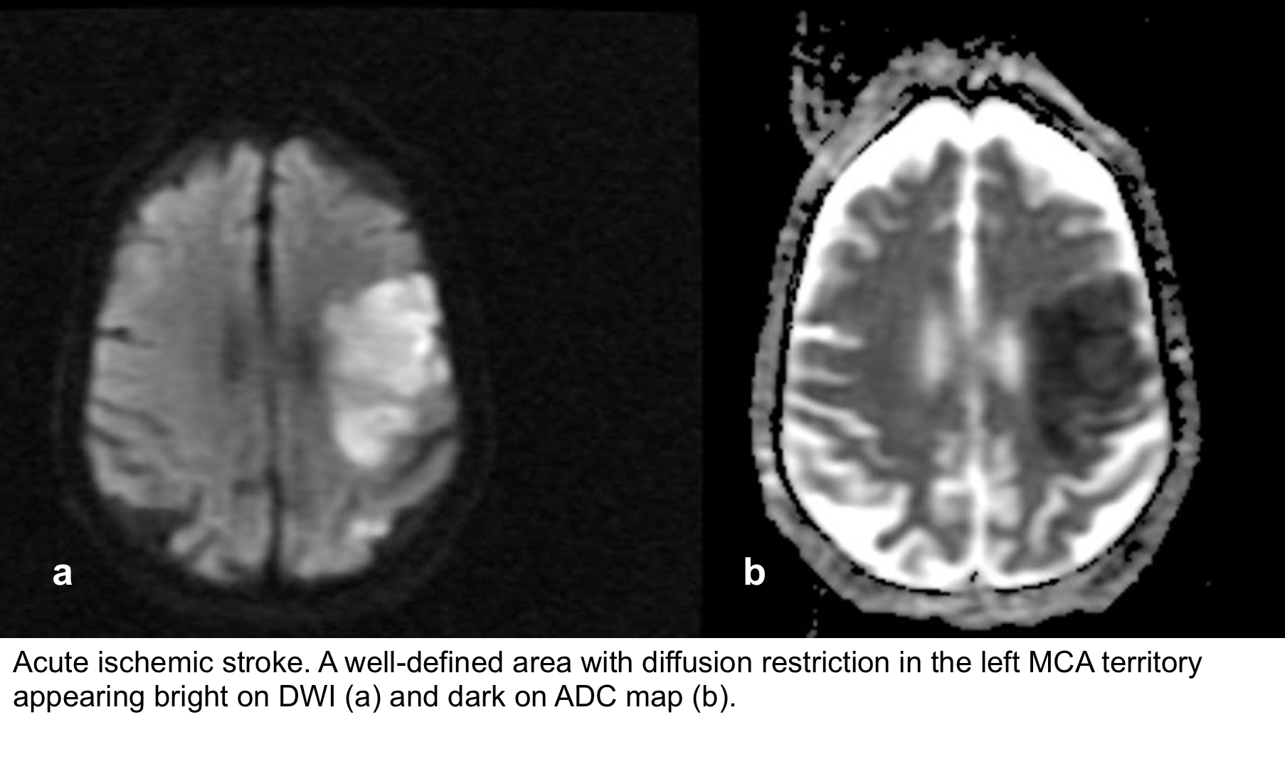
- Brain MRI with DWI is superior to NCCT for the detection of acute infarction 9.
- It is the diagnostic gold standard in acute cerebrovascular syndrome to differentiate TIA from infarction and non-ischemic mimics.
- Up to 30% of suspected TIA patients with clinical resolution of symptoms will show a rule-in infarction on MRI (Figure 9).
- Moreover, DWI can provide prognostic information in patients with ‘TIA’ 10 11 (more on this below).

- Diffusion-weighted imaging (DWI) is an MRI technique in which contrast within the image is based on microscopic motion of water.
-
- Thus, it is more sensitive to early changes of cytotoxic or vasogenic damage at the cellular level than traditional MRI measurements such as T1 or T2 relaxation rates.
- DWI is inherently a T2-weighted sequence.
- Structures with ↑diffusion, such as CSF, will appear dark on DWI images.
- Lesions with ↓diffusion will appear bright (hyperintense signal).
- ADC maps provide pure information on diffusion without any T2 weighting.
- Structures with ↑diffusion, such as CSF, will appear bright on ADC maps.
- Lesions with ↓diffusion will appear dark (hypointense signal).
- Failure of Na/K-ATPase
- Acute ischemic stroke (see image below).
- Necrotizing infections, e.g., HSV.
- Hypo-hyperglycemia.
- Drug-induced encephalopathies, such as methotrexate.
- Tissue vacuolization, spongiform changes
- Demyelination, dysmyelination
- Diffuse axonal injury.
- High protein concentration or increased viscosity
- Pyogenic infection
- Hemorrhage
- Dense cell packing
- Neoplasms such as lymphoma, high-grade glioma, and small-cell lung cancer metastases.
Ischemic changes on MRI

◾️Hyperacute (0-6hrs)
- DWI-ADC: Within minutes of infarction, DWI demonstrates ↑signal (bright) and ↓ADC value (dark).
- Other MRI sequences: the affected parenchyma appears normal, although changes in flow will be detected (occlusion on MRA), and the thromboembolism may be detected (e.g., on SWI). Slow or stagnant flow in vessels may also be detected as a loss of normal flow void and high signal on T2/FLAIR.
◾️Late hyperacute (6-24hrs)
- DWI-ADC: as described above.
- T2/FLAIR: Generally, after 6 hours, a high T2 signal will be detected, initially more easily seen on FLAIR than conventional T2.
- T1: hypointensity is only seen after 16 hours and persists.
◾️Acute (6-72hrs)
- DWI-ADC: the infarcted parenchyma continues to demonstrate ↑DWI signal (bright) and ↓ADC signal (dark).
- T2/FLAIR: remains hyperintense on T2 and FLAIR, with the T2 signal progressively increasing during the first 5 days.
- T1 signal remains low.
◾️Subacute (3 days-21 days)
- DWI-ADC: DWI remains elevated (bright) due to a persistent high T2/FLAIR signal (T2 shine-through). In contrast, ADC demonstrates pseudonormalization (becomes less dark), typically occurring between 5-14 days.
- T2 remains high, and the T1 signal remains low.
- Gadolinium-based MRI: After day 5, the cortex usually demonstrates contrast enhancement on T1 C+.
- Less common patterns of enhancement include arterial enhancement, encountered in approximately half of strokes and becomes evident after 3 days, and meningeal enhancement, which is uncommon and is usually seen between 2 and 6 days.
◾️Chronic (beyond 3 weeks)
- DWI-ADC: DWI becomes less intense and ultimately normalizes (isointense). The ADC becomes less dark and ultimately shows increased diffusivity (bright).
- The T2 signal remains high, and the T1 signal remains low.
- Gadolinium-based MRI: Cortical contrast enhancement usually persists for 2 to 4 months. Importantly, if parenchymal enhancement persists for more than 12 weeks, the presence of an underlying lesion should be considered.
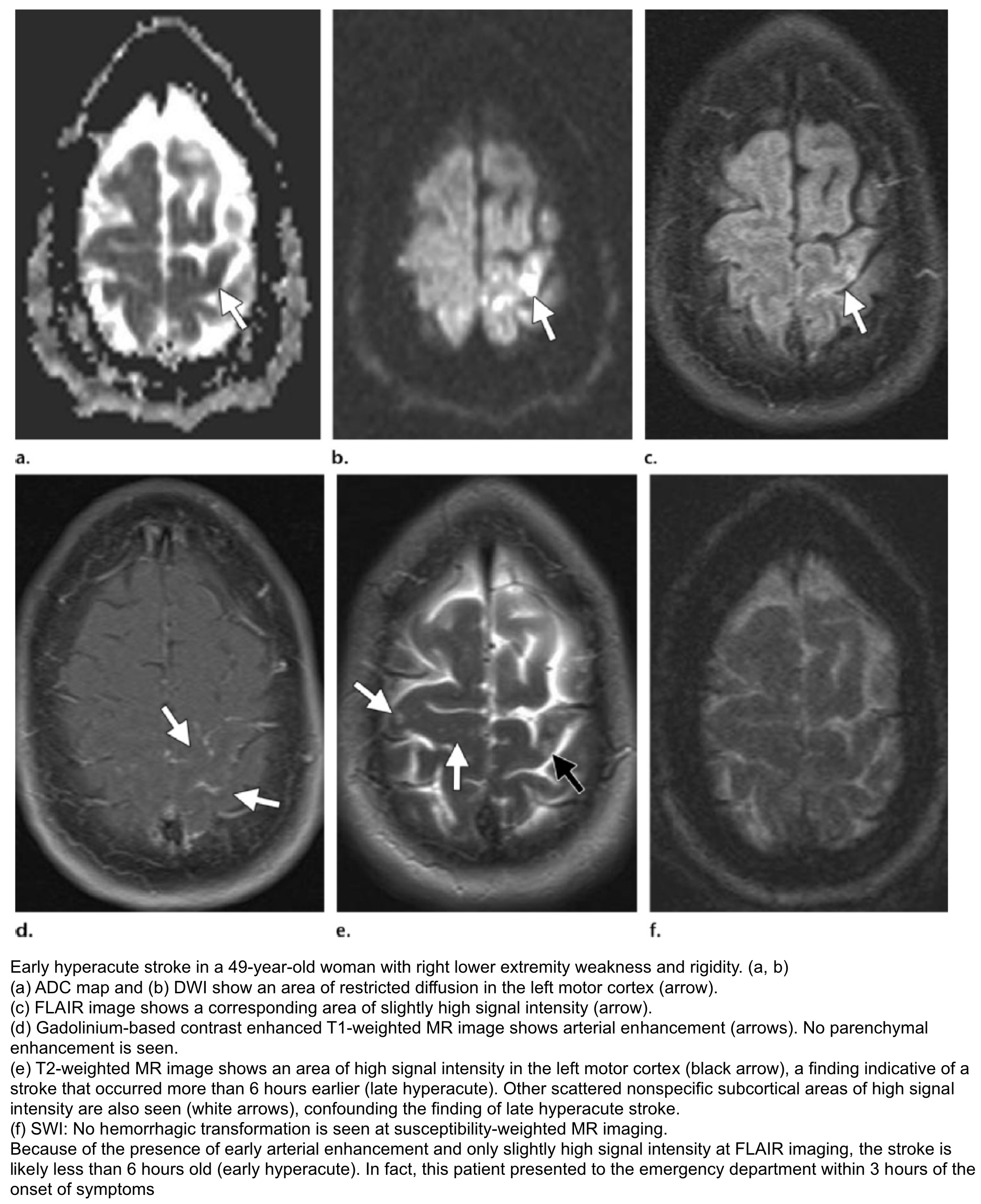
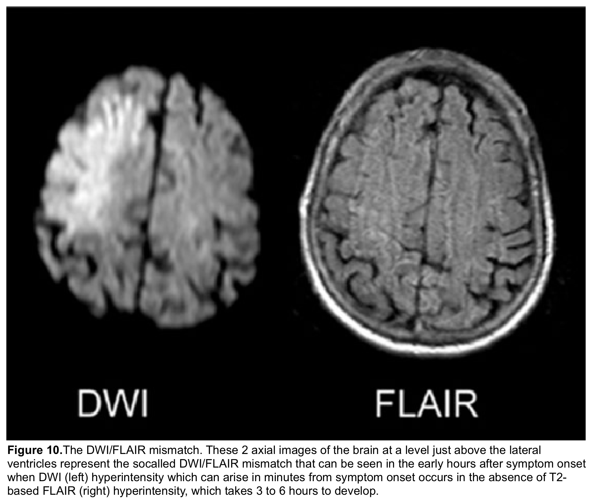
- It refers to evidence of a hyperintense lesion on DWI consistent with acute infarction, but no corresponding signal abnormality on the FLAIR images (Figure 10).
- This mismatch indicates that the stroke is relatively acute (i.e., within 4.5 h), since insufficient time has passed for the development of a hyperintense signal on FLAIR, a sign of vasogenic edema.
- In some trials, this DWI-FLAIR mismatch has been used to select patients for treatment with IVT when the time of stroke onset is unwitnessed or unknown 12.
- SWI is particularly sensitive to compounds that distort the local magnetic field. Therefore, it’s useful in detecting blood products, calcium, etc.
- SWI is the most sensitive sequence for depicting hemorrhagic transformation in patients with ischemic stroke.
- Hemorrhagic transformation demonstrates a spectrum of findings ranging from small petechial areas of microbleeding to large parenchymal hematomas.
- Micro bleeding is present in one-half to the majority of patients with ischemic stroke and is seen around 48 hours after the onset of symptoms, and is not associated with worse outcomes.
- Guidelines state that the presence of <5 areas of microbleeding on initial MR images does not contraindicate thrombolysis because they are not associated with increased adverse outcomes.
- Micro bleeding is present in one-half to the majority of patients with ischemic stroke and is seen around 48 hours after the onset of symptoms, and is not associated with worse outcomes.
- Parenchymal hematoma is a rarer type of hemorrhagic transformation that results from vessel wall rupture caused by high reperfusion pressure.
- It is more common with cardioembolic events, is associated with hyperglycemia, most commonly occurs in the basal ganglia, and confers a much worse prognosis.
- Hemorrhagic transformation demonstrates a spectrum of findings ranging from small petechial areas of microbleeding to large parenchymal hematomas.
🩸Hemorrhagic transformation is rare in the first 12 hours after stroke onset (the hyperacute stage), particularly within the first 6 hours. When it occurs, it is usually within the first 24–48 hours and, in almost all cases, is present 4–5 days after stroke.

Vascular imaging: CTA/MRA
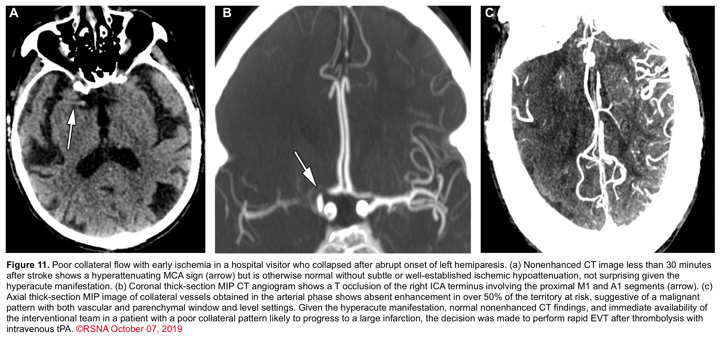
With the advent of endovascular therapies (EVT), identifying the presence of intracranial LVO is important for therapeutic decisions. CTA or MRA can detect these lesions 7 (Figure 11).
- If a patient is a possible candidate for EVT, vascular imaging is recommended concurrently with the initial head CT; however, these additional studies should not delay thrombolytic administration *.
- Practically speaking, if the clinical suspicion is high and the resources are available, neurology and pharmacy can accompany the patient to the scanner, mix tPA, recheck blood pressure, and administer thrombolysis as soon as the head CT rules out a bleed. Then, while tPA is running, proceed with the CTA of the head and neck.
- It is important to realize that in patients with no history of renal insufficiency, it is not necessary to have a serum creatinine result before performing contrasted studies for stroke, because these studies are not associated with a significantly increased risk of acute kidney injury *.
- Primary Targets (Acute Intervention)
- Large Vessel Occlusion (LVO): Presence and location of clot in ICA terminus, M1 (proximal MCA), basilar artery, or, less commonly M2, vertebral, or ACA *.
- Clot morphology: Abrupt cut-off (suggests embolus) vs. tapered/irregular (suggests in-situ atherosclerosis)
- Clot burden: Length of thrombus, presence of residual flow
- Tandem occlusion: Simultaneous cervical ICA stenosis/dissection + intracranial MCA occlusion (requires combined approach) *.
- Access & Procedural Planning
- Aortic arch anatomy: Arch type (I, II, III), bovine arch, great vessel origins (brachiocephalic, left common carotid, left subclavian) – determines catheter access difficulty *.
- Vessel tortuosity: Severe looping or kinking that may impede catheter navigation
- Aortic arch atheroma: Large or mobile plaque that could embolize during catheter manipulation.
- Collateral Assessment (Prognostic)
- Collateral flow grade: Pial collaterals filling the ischemic territory (assesses chance of good outcome and guides patient selection, especially in late window) *.
- Alternative Diagnoses (Exclude Non-Stroke Pathology)
- Arterial dissection: Tapered, flame-shaped occlusion, intimal flap, pseudoaneurysm, or intramural hematoma (look in cervical ICA and vertebral arteries)
- Vasculitis / RCVS: Vessel beading, alternating stenosis and dilatation
- Reversible cerebral vasoconstriction syndrome (RCVS): String of beads appearance
- Cerebral venous thrombosis (CVT): Empty delta sign on CTV (if venous phase acquired)
- Incidental Findings
- Aneurysm (unruptured) – may alter management (anticoagulation/antiplatelet decisions)
- Arteriovenous malformation (AVM) or other vascular malformation
- Tumor (primary or metastatic)
- Old infarcts or chronic white matter disease
- If resources are available and patients meet the general criteria for EVT, CTA should be performed for:
-
- All patients presenting within 0-4.5h of LSN.
- For patients presenting within 4.5 – 24h of LSN and NIHSS ≥6 or VAN positive or true LVO stroke syndrome described above.
💡In patients with disabling AIS, ask for the correct CTA protocol. This is explained in the following table *.
| Feature | CODE STROKE (Disabling Stroke) | URGENT CT/CTA (Non-Disabling Stroke) |
|---|---|---|
| Primary goal | Rule out hemorrhage → identify LVO → acute reperfusion (IVT/EVT) | Identify etiology (carotid stenosis, dissection, AF, etc.) for secondary prevention |
| Patient presentation | Unable to walk, talk, or perform basic ADLs Disabling deficit |
Mild symptoms (e.g., facial droop, mild sensory loss, ataxia without disability) Non-disabling deficit |
| Timeframe | STAT – within 20-25 minutes of arrival | Urgent – within 24-48 hours |
| First imaging study | NCCT – to exclude hemorrhage (STAT) | MRI (preferred) or CT if MRI not available |
| Vascular imaging | CTA head and neck – immediately after NCCT | CTA or MRA – within 24-48 hours |
| CTA coverage | Aortic arch to vertex Why? EVT planning requires: • Evaluate aortic arch anatomy (bovine arch, type I/II/III) for catheter access • Identify tandem lesions (carotid dissection/stenosis + intracranial clot) • Assess great vessel origins (brachiocephalic, subclavian, vertebral) • Plan optimal vascular access route for thrombectomy |
Carina to circle of Willis Why? Focused evaluation of: • Carotid bifurcation stenosis (for CEA/CAS candidacy) • Intracranial stenosis • No need for arch anatomy (acute intervention not planned) |
| CT Perfusion (CTP) | Required for late window (6-24h) to assess penumbra (core <70 mL, mismatch ratio >1.8, mismatch volume >15 mL) | Not usually required (unless LVO unexpectedly found) |
| What radiologist looks for | • Hemorrhage? (yes/no) • LVO? (ICA terminus, M1, basilar) • Mismatch? (for late-window EVT) • Abrupt cut-off (embolic) • Tandem lesions (cervical + intracranial) |
• Stenosis ≥50%? (carotid, intracranial) • Plaque ulceration, dissection flap • Old infarcts, tumor, atrophy • Tapered, irregular stenosis (atherosclerosis) |
| IV Thrombolysis (IVT) | ✅ Indicated if disabling deficit, ≤4.5h (or extended window with mismatch) – Class 1 | ❌ NOT indicated for non-disabling deficits – Class 3 (DAPT preferred) |
| EVT (Thrombectomy) | ✅ Indicated if LVO within 24h (with mismatch if 6-24h) – Class 1 | ❌ Not indicated (unless CTA unexpectedly shows LVO → upgrade to CODE STROKE) |
| Antiplatelet therapy | Aspirin started 24h after IVT (if no hemorrhage) | DAPT (if minor stroke/TIA, ABCD² ≥4) or aspirin monotherapy – start early |
| Anticoagulation (for AF) | Delayed 24h after IVT; early (≤4 days) reasonable with DOAC (Class 2a) | Started early (≤4 days) if no hemorrhage – DOAC preferred |
| Disposition | ICU or stroke unit – prepare for IVT/EVT | Ward or rapid-access TIA clinic – outpatient follow-up possible for low-risk |
1. EVT access planning: Neurointerventionalists need to see the aortic arch type (I, II, III), great vessel origins (brachiocephalic, left common carotid, left subclavian), and any arch atheroma or bovine variants that may affect catheter navigation.
2. Tandem lesion detection: Many LVOs are caused by a proximal cervical ICA stenosis or dissection (tandem lesion). Full coverage identifies both the proximal cause and the distal clot, guiding combined treatment (stenting + thrombectomy).
3. Vertebral and basilar assessment: For posterior circulation strokes, arch coverage captures vertebral artery origins (often from subclavian), which may be occluded or dissected.
4. Collateral pathway evaluation: Complete arch and neck imaging helps assess potential collateral routes (e.g., external carotid to ophthalmic, posterior communicating artery).
5. No need for separate studies: One acquisition provides all information needed for EVT triage, avoiding delays.
➡️ In contrast, urgent CTA for non-disabling stroke only needs coverage from carina to circle of Willis (carotid bifurcation focus) because acute intervention is not planned.
✅ Key takeaway: CODE STROKE (disabling) – STAT NCCT → CTA (arch to vertex) → CTP if 6-24h → goal is acute reperfusion (IVT/EVT). URGENT (non-disabling) – MRI + CTA within 24-48 hours (carina to circle of Willis) → goal is etiology detection for secondary prevention. IVT is NOT recommended for non-disabling deficits (Class 3).
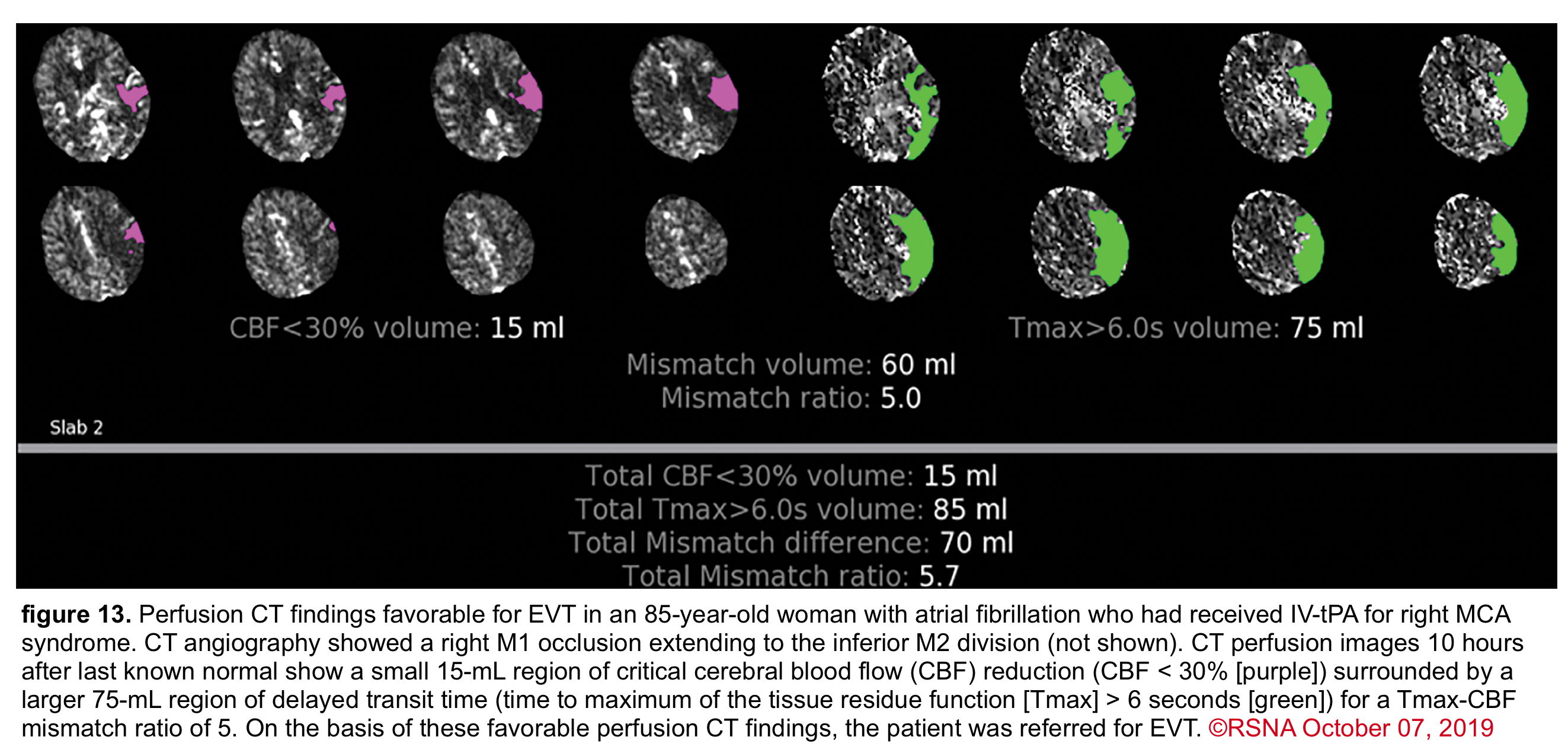
Perfusion Study
In acute ischemic stroke, the area of irreversible brain infarct (core) is surrounded by ischemic tissue (penumbra) that may potentially be salvageable (Figure 12), regardless of the time of onset of symptoms.
- The infarct core refers to tissue that has already been damaged. Even if the vessel could be immediately opened, the core infarct would not recover.
- Radiological definition of core infarct is based on the development of cytotoxic edema, reflective of neuronal cells swelling.
- This cytotoxic edema causes a diffusion restriction on MRI, which is the reference standard for defining the core infarct.
- Radiological definition of core infarct is based on the development of cytotoxic edema, reflective of neuronal cells swelling.
- Ischemic penumbra is the tissue that surrounds the core infarct. The ischemic penumbra is malperfused and nonfunctional, but the tissue is still potentially viable if the blood supply can be restored. The ischemic penumbra is often maintained by a thin and slow blood flow supplied via collateral circulation.
- The entire purpose of revascularization is to revive the ischemic penumbra. Alternatively, if the ischemic penumbra is small, then there is little more tissue to salvage; the stroke has already completed.

📍Perfusion study (Multimodal CT or MRI) can provide crucial information about the volume of tissue that is irreversibly damaged (infarction core) and the volume of tissue that is critically hypoperfused but potentially salvageable with reperfusion (ischemic penumbra) 7. This will guide further therapy for patients who fall outside the time ranges for thrombolysis or where the time of symptom onset is unclear.
- The mantra of stroke is that “time is brain.” However, this is only partially true because different patients progress at different speeds over time.
- The pace of ischemic penumbra necrosis varies widely among patients and depends on the degree of collateral circulation.
- Some patients may rapidly complete an infarction within hours, while others may continue to have a large ischemic penumbra for many hours. The latter group may remain a good candidate for revascularization therapy beyond the traditional IVT window.
- 💡Therefore, the 4.5 hours could have an entirely different meaning among different groups of patients.
◾️Moving from “time-threshold” to “tissue-threshold.”
- With the advent of modern neuroimaging, it is possible to rapidly determine the amount of salvageable tissue. This may largely replace time cutoffs when considering candidacy for interventions.
◾️CT-perfusion parameters
- Cerebral blood flow (CBF) and time to maximum enhancement are among the most accurate values for use in acute stroke evaluation.
- Cerebral blood flow < 30% is used to identify the core infarct.
- Maximal transit time (Tmax) relates to the maximal time blood spends in a given region.
- Elevated maximal transit time using a cutoff of 6 seconds identifies both the core infarct and ischemic penumbra.
💡The key issue is the degree of mismatch between the two images:
- If the two images match up, then the volume of ischemic penumbra is small (completed infarct).
- If the ischemic core is much smaller, then the volume of ischemic penumbra must be large (implying significant salvageable brain tissue). The mismatch ratio of penumbra/core (ratios >1.8 suggest benefit from EVT).
Imaging Decision Making in AIS Management
Brain imaging serves two critical purposes in acute ischemic stroke:
- Exclude hemorrhage (to determine eligibility for IV thrombolysis)
- Identify large vessel occlusion (LVO) and assess ischemic core vs. penumbra (to determine eligibility for EVT) *.
The choice of imaging modality and the factors guiding interpretation depend largely on the time window and the treatment being considered *.
| Decision Factor | Clinical Question | Imaging Modality | Decision Implication |
|---|---|---|---|
| 1. Intracranial hemorrhage | Is there any acute blood? | NCCT (first-line) or MRI (GRE/SWI) | Absolute exclusion for IVT and EVT if acute hemorrhage present |
| 2. Early ischemic changes / hypodensity | Is there clear hypodensity > contralateral white matter? | NCCT (ASPECTS score) | Clear hypodensity → Absolute exclusion for IVT. Low ASPECTS (0-2) reduces EVT benefit magnitude |
| 3. Large vessel occlusion (LVO) | Is there an accessible clot (ICA, M1, basilar artery)? | CTA or MRA (cervical + intracranial) | Required for EVT eligibility. Do not delay IVT to obtain CTA |
| 4. Ischemic core volume | How much brain tissue is already irreversibly injured? | NCCT (hypodensity), CTP (rCBF <30%), or DWI-MRI | Small core (<70 mL) favors treatment. Large core (70-100+ mL) reduces benefit but does not exclude EVT (Class 2a for ASPECTS 0-5) |
| 5. Penumbra (salvageable tissue) volume | How much brain is at risk but potentially recoverable? | CTP (Tmax >6 sec) or MR PWI | Large penumbra + small core → ideal candidate for late-window EVT |
| 6. Perfusion-core mismatch ratio | Is salvageable tissue >1.8 × core volume? | CTP or MR DWI/PWI with automated software | Required for late-window (6-24h) EVT per DAWN/DEFUSE-3 criteria |
| 7. Collateral perfusion | How robust is alternative blood flow to the ischemic territory? | Multiphase CTA or single-phase CTA | Good collaterals → slower core growth → more likely to benefit in late window. Poor collaterals → rapid penumbra collapse |
| 8. Time window | Is the patient in early (0-6h) or late (6-24h) window? | Clinical history + imaging | Early window (0-6h): Advanced imaging optional. Late window (6-24h): Advanced imaging required to demonstrate mismatch |
| 9. Unknown time of onset / wake-up stroke | Can we estimate stroke age and identify salvageable tissue? | MRI DWI-FLAIR mismatch (for IVT) or CTP/MR PWI (for EVT) | DWI-FLAIR mismatch → IVT eligible up to 4.5h from awakening. Perfusion mismatch → EVT eligible 6-24h from last known well |
| 10. Posterior circulation stroke | Is there basilar artery occlusion or posterior circulation ischemia? | CTA (basilar artery), PC-ASPECTS on NCCT or DWI | PC-ASPECTS ≥6 favors EVT for basilar occlusion within 24h. DWI more sensitive than NCCT for posterior fossa |
🕒 Imaging Decision Tree by Time Window:
• 0 – 4.5 hours (IVT window): NCCT to exclude hemorrhage → give IVT if disabling deficits. Do NOT delay for CTA or perfusion.
• 0 – 6 hours (early EVT window): NCCT + CTA to identify LVO. Advanced perfusion imaging is optional (not required).
• 6 – 24 hours (late EVT window): NCCT + CTA + required advanced perfusion imaging (CTP or MR DWI/PWI) to demonstrate small core + large penumbra mismatch.
• Wake-up / unknown onset: MRI DWI-FLAIR mismatch for IVT eligibility; CTP or MR PWI mismatch for EVT eligibility.
🧬 Core Biological Principle (2026 Guideline):
Ischemic stroke comprises three tissue compartments: irreversible core, salvageable penumbra, and normal tissue. Reperfusion therapies aim to rescue the penumbra. Robust collaterals sustain penumbral viability into the late window, whereas poor collaterals accelerate its collapse. Advanced imaging that reveals a small core with a large penumbra identifies patients most likely to achieve favorable functional outcomes—even beyond 6 hours.
✅ Abbreviations: NCCT = non-contrast CT; CTA = CT angiography; CTP = CT perfusion; MRI = magnetic resonance imaging; DWI = diffusion-weighted imaging; PWI = perfusion-weighted imaging; FLAIR = fluid-attenuated inversion recovery; ASPECTS = Alberta Stroke Program Early CT Score; PC-ASPECTS = posterior circulation ASPECTS; rCBF = regional cerebral blood flow; LVO = large vessel occlusion; IVT = intravenous thrombolysis; EVT = endovascular thrombectomy.
⚠️ Critical rule: For patients within 4.5 hours with disabling deficits, do not delay IVT to obtain CTA, CTP, or MRI. For late-window EVT (6-24h), advanced perfusion imaging is mandatory to demonstrate salvageable tissue.
Disabling AIS Management
General supportive early management
- All patients with suspected acute stroke should be assessed immediately upon arrival for airway compromise 6. Patients who are unable to clear oral secretions or maintain airway stability should be immediately intubated ( neurocritical care intubation).
- Provide supplemental O2 if oxygen saturation is <94%. Supplemental O2 is not recommended in non-hypoxic patients with ‘AIS’ 6.
All patients with suspected acute stroke should be assessed immediately upon arrival for hemodynamic stability 6.
- ⚠️Hypotension
- Hypotension with evidence of poor perfusion (shock state) can mimic stroke, especially in elderly patients, and should be appropriately managed (before imaging).
- Avoid hypotension at all costs, as it accelerates penumbral infarction *.
- Several studies have found an association between hypotension and worse neurological outcomes *.
- Evaluate for the etiology of hypotension and treat any causes (e.g., hypovolemia).
- Discontinue any antihypertensives.
- The use of vasopressors here is not evidence-based, but could be reasonable in specific patient scenarios (e.g., the perfusion-dependent patient whose neurological exam worsens due to hypotension, in which vasopressors are started, and this improves the neurological exam).
- Several studies have found an association between hypotension and worse neurological outcomes *.
- Hypertension management
- BP for patients not receiving any intervention
- Hypertension is a normal, physiologic response that improves brain perfusion. This should generally be left alone for the first ~24-48 hours. Over time, BP will generally decrease on its own. Reduction in blood pressure is indicated only if there is *:
- Hypertensive emergency with target organ damage (e.g., myocardial ischemia, hypertensive nephropathy, pulmonary edema). Hypertensive emergency is discussed further here.
- BP is >220/120 mm Hg. However, even in this situation, the benefit of treating hypertension is uncertain *.
- If blood pressure reduction is needed, this should be gentle (e.g., ~15% reduction during the first 24 hours, unless there is target organ damage) *.
- Hypertension is a normal, physiologic response that improves brain perfusion. This should generally be left alone for the first ~24-48 hours. Over time, BP will generally decrease on its own. Reduction in blood pressure is indicated only if there is *:
- BP for patients receiving thrombolysis alone (Figure 18 below).
- BP control for patients receiving endovascular therapy *
- Before thrombectomy:
- Maintain a flat position to improve perfusion.
- It is reasonable to maintain BP <185/110 *.
- For patients undergoing intubation before endovascular therapy, avoid hypotension. Even small reductions in blood pressure (e.g., >10% decrease) may correlate with worse outcomes.
- After thrombectomy:
- Most guidelines recommend <180/105 (similar to post-thrombolysis patients) *.
- A reasonable goal may be SBP 140-180 mm *. This also aligns with the BP target for patients who received thrombolysis, which is convenient since many of these patients also received thrombolysis.
- RCTs demonstrated worse outcomes with more intensive BP control (targeting SBP <140) *.
- Before thrombectomy:
- 💡The approach to blood pressure management in ‘AIS’ is inherently different from the approach in acute hemorrhagic stroke (see here).
- For this reason, a neuroimaging study (CT or MRI) is critical to help guide blood pressure therapy in patients with acute stroke.
- BP for patients not receiving any intervention
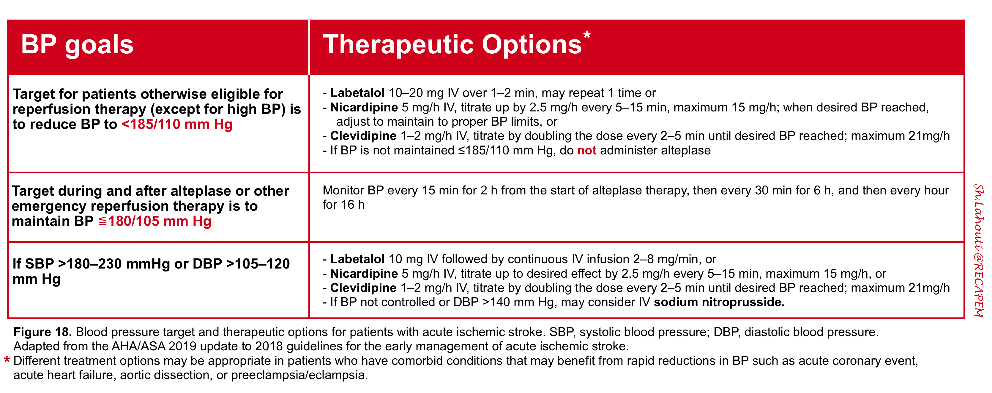
- IV Access
- Obtain peripheral intravenous (IV) access and avoid unnecessary lines and ABG, since minor vascular trauma in patients with ischemic CVA who are deemed to be candidates for thrombolysis may become a real problem.
- 🔬Initiate labwork
- STAT fingerstick glucose.
- Complete blood count.
- Electrolytes, including Ca/Mg/Phos.
- Liver function tests.
- Coagulation studies:
- Generally, PT, PTT, and fibrinogen.
- Anti-Xa (or dedicated apixaban level) for patients on oral Xa inhibitors.
- Blood cultures x2 if concern for endocarditis (e.g., fever or history of IV drug use).
- Pregnancy test as appropriate
- Perform a focused neurological exam and obtain a point-of-care glucose.
- The focused exam is structured around relevant data gathered during the medical history and is tailored to the differential diagnosis. The examiner should be focused on determining whether (1) there is a lesion and (2) where the lesion is localized *.
- Baseline ECG assessment is recommended in patients presenting with “AIS” but should not delay initiation of IVT in selected patients *.
- This is to protect against aspiration.
- Dysphagia is common in patients with acute stroke and is associated with the development of aspiration pneumonia.
- Dysphagia related to stroke is more precisely characterized by oropharyngeal dysphagia, defined by swallowing impairment of the upper digestive tract.
- Cranial nerves involved in swallowing include: sensory components of CN V, IX, X, and motor components of CN V, VII, X, XI, and XII.
- All patients should receive a bedside swallowing screening examination before initiation of a diet (e.g., supervised ability to drink a glass of water by a bedside nurse or speech and language therapist).
- The test is passed if the patient can drink the entire volume of water (in a single or sequential swallows) without coughing, gurgling, or choking during or immediately (e.g., within one minute) after swallowing.
- It is important to keep the patient ‘NPO’ (nothing by mouth) to protect against aspiration until the swallowing function is evaluated 6.
🕰️ Nonessential testing and procedures should not delay performing brain imaging within 25 minutes of the patient’s arrival 6.
Reperfusion Therapy
◾️The immediate goal of reperfusion treatment for ‘AIS’ is to salvage regions of the brain that are ischemic but not yet infarcted. The long-term goal is to reduce stroke-related disability and mortality. The options for reperfusion therapy are IVT and EVT.
🔑 Key information needed *
- What is the LSN time?
- What is the specific deficit (disabling or NOT)
- Thrombolytic contraindications
- Goals of care and premorbid conditions?
- Establishing goals of care: A 4-part approach
- Emphasize the critical nature of time and why this discussion matters immediately.
- Describe the situation and the potential paths (IV Thrombolysis/EVT versus conservative management), covering best and worst possible results and bleeding dangers for each option.
- Align recommendations with what matters most to the patient (include 1. home independence, 2. return to work, 3. acceptable long-term disability)
- Provide your straightforward, easy-to-understand recommendation.
- Evaluating Premorbid Condition
- Premorbid condition refers to the patient’s level of function, independence, and overall health status before the acute stroke occurred.
- Assessing this is critical because it influences treatment decisions, prognosis, and goals of care.
- Evaluation
- Two-question premorbid screen:
- Where does the patient live? (independent home vs assisted vs long-term care)
- How do they mobilize? (independent vs aid vs bed-bound). This rapidly informs whether aggressive therapy aligns with baseline quality of life.
-
Modified Rankin Scale (mRS) for Neurologic Disability 🧮MDCalc
- Two-question premorbid screen:
- Premorbid condition refers to the patient’s level of function, independence, and overall health status before the acute stroke occurred.
- Establishing goals of care: A 4-part approach
| Intravenous Thrombolytic Therapy (IVT) |
Intravenous thrombolysis (IVT) is a cornerstone of early reperfusion therapy for eligible patients with acute ischemic stroke (AIS). The 2026 AHA/ASA Guideline continues to strongly recommend IVT for patients with disabling neurological deficits who present within 4.5 hours of last known well, without evidence of intracranial hemorrhage on non-contrast CT *.
◾️The Evidence for Systemic Thrombolytics in Ischemic Stroke Management
- Most evidence supports thrombolytic administration ≤ 4.5hr from onset (window opened from 3hr in 2008 to 4.5hr)
- Improves functional outcome (modified Rankin score, NNT ~7 for good outcome if given promptly)
- Best results ≤ 90min; efficacy diminishes with time but remains present ≤ 4.5hr
- Risks:
- Symptomatic intracerebral hemorrhage 3–5%
- Door-to-needle:
- Goal is ≤ 30–60 minutes *.
◾️Two thrombolytic agents are now equally recommended: Alteplase (tPA) vs. Tenecteplase (TNK) *
- Tenecteplase (TNK):
| Feature / Aspect | Alteplase (tPA) | Tenecteplase (TNK) |
|---|---|---|
| 2026 Guideline Recommendation | Class 1, LOE A Recommended |
Class 1, LOE A Recommended (non‑inferior) |
| Dosage | 0.9 mg/kg body weight (maximum 90 mg) |
0.25 mg/kg body weight (maximum 25 mg) |
| Administration | Bolus (10% of dose) + 60‑minute IV infusion | Single IV bolus over 5–10 seconds Simpler, faster workflow |
| Time window | Within 4.5 hours of symptom onset or last known well (standard window) | |
| Efficacy | Reference standard – improves functional outcomes | Non‑inferior to alteplase; multiple phase 3 RCTs (ATTEST‑2, AcT, TRACE‑2, TASTE) show similar functional outcomes (mRS 0‑1 at 90 days) |
| Safety | Well‑established; risk of sICH ~2‑7% in eligible patients | Similar safety profile; no significant difference in symptomatic intracranial hemorrhage (sICH) or mortality |
| ⚠️ Dose NOT recommended | — | Tenecteplase 0.4 mg/kg is NOT recommended (Class 3, No Benefit). Higher dose provides no added benefit and may increase harm (NOR‑TEST 2). |
| Practical advantage | Familiar, widely available, extensive real‑world experience | Single bolus reduces door‑to‑needle time, eliminates infusion pump errors, simplifies transfer cases |
| Bridging before EVT | Standard bridging therapy; proven in multiple trials | Acceptable alternative (EXTEND‑IA TNK, ACT). No clear difference in recanalization rates vs alteplase prior to thrombectomy. |
| Extended window (4.5‑24h) | Used in EXTEND, ECASS‑4 (perfusion selection) | Tenecteplase studied in TRACE‑III, TIMELESS; benefit mainly if EVT not rapidly available. No benefit over placebo when rapid EVT performed (TIMELESS). |
• Both agents are recommended within 4.5 hours for eligible patients with disabling deficits.
• Tenecteplase 0.25 mg/kg is non‑inferior to alteplase; the single‑bolus regimen may simplify logistics.
• Higher dose tenecteplase (0.4 mg/kg) should not be used.
• Choice depends on hospital protocol, cost, availability, and clinician preference — both are endorsed as first‑line options.
Decision making
- All adult patients with a clinical diagnosis of ‘AIS’ should be rapidly screened for treatment with IVT.
- Simultaneously, patients with suspected ‘AIS’ involving the anterior circulation should be evaluated for EVT.
✅ Inclusion criteria for Acute Ischemic Stroke *
| Parameter | Specification / Notes (2026 Guideline) |
|---|---|
| Diagnosis | Acute ischemic stroke with disabling neurological deficits (functional assessment: inability to perform basic ADLs such as bathing, walking, toileting, eating; or inability to return to work). NIHSS alone is not decisive. |
| Time window (standard) | Within 4.5 hours from symptom onset or last known well. |
| Time window (extended) | 4.5–9 hours from last known well OR wake-up stroke within 9 hours from midpoint of sleep, with advanced imaging mismatch (CTP or MRI DWI-FLAIR) showing salvageable penumbra (Class 2a). |
| Imaging requirement | No intracranial hemorrhage on NCCT or MRI. Early ischemic changes (including low ASPECTS) are NOT a contraindication. |
| Stroke severity | Any NIHSS score, as long as deficits are disabling (see above). |
| Blood pressure | SBP ≤185 mm Hg AND DBP ≤110 mm Hg after correction (if needed) before IVT initiation. |
| Glucose & coagulation | Blood glucose >50 mg/dL (correct hypoglycemia first). Do NOT delay IVT for coagulation testing unless clinical suspicion of coagulopathy. |
| Age | Adults ≥18 years. Pediatric patients (28 days – 18 years) may be considered (Class 2b, safety known but efficacy uncertain). |
| Treatment speed | Faster treatment improves outcomes. Do NOT delay for CTA, CT perfusion, or advanced imaging; do NOT wait for coagulation tests if no suspicion of abnormality. |
🚫Contraindications to IV Thrombolytics in Ischemic Stroke Management Update 2025 * 👇
| Condition / Finding | Specific criteria (2026 AHA/ASA) |
|---|---|
| Intracranial hemorrhage | Acute hemorrhage visible on CT or MRI. |
| Extensive clear hypodensity | Regions of clear hypodensity on NCCT greater than density of contralateral white matter, and responsible for clinical symptoms. |
| Recent severe TBI | Moderate/severe traumatic brain injury within <14 days with unconsciousness >30 min, GCS <13, or hemorrhage/contusion/skull fracture on imaging. |
| Recent CNS surgery | Intracranial or spinal surgery within <14 days. |
| Spinal cord injury | Within <3 months. |
| Active bleeding / coagulopathy | Active internal bleeding; platelets <100,000/mm³, INR >1.7, aPTT >40s, PT >15s (if available). |
| Aortic arch dissection | Known or strongly suspected aortic dissection. |
| Infective endocarditis | Suspected or confirmed infective endocarditis. |
| Intra-axial neoplasm | Intraparenchymal brain tumor. |
| ARIA (amyloid-related imaging abnormalities) | Patients receiving amyloid immunotherapy with known ARIA. |
| Category / Condition | Guideline recommendation & clinical nuance |
|---|---|
| Recent DOAC use (<48h) | Safety unknown. May be considered after individual risk/benefit analysis (relative, not absolute). Emerging data, but definitive trials needed. |
| Prior intracranial hemorrhage (ICH) | History of ICH (especially cerebral amyloid angiopathy). Individualized decision, potential increased risk. |
| Recent ischemic stroke (<3 months) | Higher hemorrhagic conversion risk. Weigh benefit against risk; case-by-case. |
| Pre‑existing disability (mRS 3–4) | Benefit uncertain; consider functional goals, but IVT may still be considered after shared decision‑making. |
| High burden cerebral microbleeds (>10 CMBs) | If known >10 microbleeds on MRI, usefulness uncertain. Do NOT delay IVT to obtain MRI for screening. |
| Recent major surgery (<10 days) | Major non‑CNS surgery within 10 days; surgical consultation advised. |
| Recent major trauma (14d – 3m) | Non‑CNS major trauma within 14 days to 3 months. |
| Recent GI/GU bleeding (<21 days) | Gastrointestinal or genitourinary bleeding within 21 days. |
| Arterial puncture of non‑compressible vessel (<7d) | E.g., subclavian line; consider risk. |
| Intracranial arterial dissection | Safety unknown; careful individual assessment. |
| Unruptured intracranial vascular malformation | Potential hemorrhagic risk; weigh against stroke severity. |
| Intracardiac thrombus (LA/LV) | Cardiology consultation recommended. |
| Acute pericarditis / recent STEMI (<3 months) | Emergent cardiology consultation before IVT. |
| Pregnancy / postpartum | May be considered for moderate/severe disabling stroke with obstetrical consultation. |
| Systemic active malignancy | Oncology input to balance bleeding risk vs potential benefit. |
| ❌ CLASS 3: NO BENEFIT (Non‑disabling deficits) | IVT is NOT recommended for patients with mild non‑disabling deficits (e.g., isolated sensory syndrome, minor weakness that does not impair ADLs). Dual antiplatelet therapy (DAPT) is preferred in this subgroup (Class 3, No Benefit). |
Consent For TPA/TNK
- Shared decision-making for IVT should include:
- Diagnosis of ischemic stroke with disabling deficit
- Absolute benefit (e.g., 6 more good outcomes per 100 treated)
- Absolute risk of sICH (e.g., 2 per 100)
- Urgency of early treatment
- The alternative of no treatment.
- 💡If the patient cannot participate and no proxy is immediately available, proceed with treatment — delay is more harmful than proceeding without consent *.
Administration
Dose: 0.9 mg/kg (max 90 mg)
Bolus: 10% of total dose IV over 1 minute
Infusion: Remaining 90% over 60 minutes (infusion pump)
Max total dose: 90 mg
Recommendation: Class 1, LOE A
Dose: 0.25 mg/kg (max 25 mg)
Administration: Single IV bolus over 5–10 seconds (no infusion pump)
Infusion: None – single push
Max total dose: 25 mg
Recommendation: Class 1, LOE A (equally recommended)
| Weight (kg) | TNK Dose (mg) | Volume (mL) |
|---|---|---|
| <60 kg | 15 mg | 3 mL |
| 60 – <70 kg | 17.5 mg | 3.5 mL |
| 70 – <80 kg | 20 mg | 4 mL |
| 80 – <90 kg | 22.5 mg | 4.5 mL |
| ≥90 kg | 25 mg | 5 mL |
- Target (first 24h): SBP <180 mm Hg AND DBP <105 mm Hg (Class 1, LOE B-R)
- Intensive SBP lowering to <140 mm Hg: NOT recommended (Class 3, No Benefit) – does not improve outcome and may cause harm.
- If BP exceeds target: increase monitoring, administer IV antihypertensive (labetalol, nicardipine).
Next 6 hours (hours 2–8): every 30 minutes
Next 16 hours (hours 8–24): every 1 hour
| Complication | Immediate action |
|---|---|
| Symptomatic ICH (sICH) | Stop infusion → emergency CT → cryoprecipitate (10 U) / tranexamic acid / aminocaproic acid → neurosurgery consult |
| Orolingual angioedema | Stop infusion, maintain airway, methylprednisolone 125 mg IV + diphenhydramine 50 mg IV + famotidine 20 mg IV; consider epinephrine/icatibant if severe |
- Delay invasive lines (nasogastric tubes, indwelling bladder catheters, intra‑arterial catheters) if safe to manage without them.
- Avoid arterial punctures if possible; if needed, use compressible sites.
- Do NOT start antiplatelet or anticoagulant therapy within the first 24 hours after IVT.
- Follow‑up imaging: Obtain CT or MRI at 24 hours before starting antiplatelet/anticoagulant agents.
| Parameter | Alteplase (tPA) | Tenecteplase (TNK) |
|---|---|---|
| Dose | 0.9 mg/kg (max 90 mg) | 0.25 mg/kg (max 25 mg) |
| Bolus | 10% over 1 min | Entire dose over 5–10 sec |
| Infusion duration | 60 min (pump) | None (single push) |
| BP target (24h) | <180/105 mmHg | |
| Intensive BP (<140) | NOT recommended (Class 3) | |
| Monitoring frequency | q15 min x2h → q30 min x6h → hourly until 24h | |
| Follow‑up imaging | CT/MRI at 24h before antiplatelets/anticoagulants | |
✅ Tenecteplase 0.25 mg/kg single bolus = equally recommended (Class 1, LOE A).
❌ Tenecteplase 0.4 mg/kg NOT recommended (Class 3, No Benefit).
❌ Intensive SBP lowering to <140 mmHg after IVT NOT recommended (Class 3).
🧠 Disabling vs non‑disabling: IVT only for disabling deficits (non‑disabling → DAPT preferred).
📉 Low‑intensity monitoring may be safe in selected mild strokes (OPTIMIST), but standard monitoring remains default.
⏱️ Time is brain: Do NOT delay thrombolysis to obtain CTA, CTP, or coagulation labs unless strongly indicated.
- 🧓Age ≥80 is NOT a contraindication – treat if otherwise eligible *.
- ⏳Time window *
- The standard IVT window remains ≤4.5 hours.
- The extended window (4.5-9h) requires advanced imaging mismatch.
- IVT may be considered up to 24h in select LVO patients who cannot get EVT
- Wake-up strokes ARE now eligible with advanced imaging.
- Beyond 24 hours: not eligible for either IVT or EVT.
- Non-disabling deficits: IVT NOT recommended (Class 3) – DAPT preferred *.
- Intensive BP lowering (<140 mmHg): NOT recommended after IVT *
- Cervical dissection is NOT a contraindication *.
- Extracranial cervical dissections: IV thrombolysis in AIS known or suspected to be associated with extracranial cervical arterial dissection is reasonably safe within 4.5 h and probably recommended.
- ‼️Note: Aortic dissection remains an absolute contraindication.
- Seizure at onset is NOT a contraindication (for IVT) if deficits are from Acute Ischemic Stroke *.
- If the deficits are due to stroke (not postictal Todd’s paresis), the patient should be treated.
- Cerebral microbleeds (CMBs)
- Do NOT delay IVT to obtain an MRI. If >10 CMBs known, usefulness uncertain (relative) *.
- Hematologic considerations 👇
In most cases, results of routine laboratory tests, including coagulation parameters and platelet count, are NOT required to proceed with IV thrombolysis. IVT should not be delayed while results are pending unless one of the following conditions is present:
- Clinical suspicion of a bleeding abnormality or thrombocytopenia
- Current or recent use of anticoagulants (heparin, warfarin, DOACs)
- Use of anticoagulants is not known
Status: Now a RELATIVE contraindication (not absolute). Individualized decision-making required.
Recommendation (2026 Guideline, Table 8, Page 50):
- “In patients with disabling symptoms and recent DOAC exposure (<48 hours) who are within the window for alteplase/tenecteplase, the safety of IV thrombolysis is unknown. Emerging but limited observational data suggest IV thrombolysis may be considered after a thorough benefit vs risk analysis on an individual basis.”
Factors to consider:
- Timing of last DOAC dose
- Renal function (DOAC accumulation)
- Stroke severity (disabling vs. non-disabling)
- Availability of endovascular thrombectomy (EVT)
- Availability of DOAC reversal agents or anti-factor Xa assays (may cause delay)
If laboratory results become available during or after treatment initiation, IVT should be discontinued if results fall outside the following ranges:
| Parameter | Threshold for discontinuation |
|---|---|
| Platelets (PLT) | < 100,000/mm³ |
| International Normalized Ratio (INR) | > 1.7 |
| Activated Partial Thromboplastin Time (aPTT) | > 40 seconds |
| Prothrombin Time (PT) | > 15 seconds |
Recommendation (Class 1, LOE B-NR):
- In otherwise eligible patients who were taking single antiplatelet therapy (SAPT) or dual antiplatelet therapy (DAPT) before the stroke, IVT is recommended.
- Despite a small increased absolute risk of symptomatic intracranial hemorrhage (sICH) of approximately 0.9-1.2%, the anticipated absolute treatment benefit (~8%) outweighs the risk.
• Do NOT delay IVT for routine labs unless clinical suspicion of coagulopathy or anticoagulant use.
• DOAC exposure <48h is now a relative contraindication – individualize decision (MAJOR 2026 UPDATE).
• Discontinue IVT if PLT <100k, INR >1.7, aPTT >40s, or PT >15s.
• Antiplatelet therapy (SAPT or DAPT) is NOT a contraindication – IVT is recommended (Class 1).
✅ Key 2026 updates reflected: DOAC exposure <48h is now a relative (not absolute) contraindication; antiplatelet therapy remains an indication FOR treatment; routine labs should not delay treatment.
Thrombolytic complications management
In patients undergoing fibrinolytic therapy, physicians should be prepared to treat potential emergent adverse effects, including bleeding complications and angioedema that may cause partial airway obstruction 6.
- Early neurological worsening
- In patients treated with thrombolytics, the most feared complication is tPA-related hemorrhage. However, other possibilities for worsening neurologic function should be considered. This include
- ⚡️Stroke-related seizure
- Stuttering lacunar infarct
- Re-occlusion of a vessel
- 🩸Symptomatic intracerebral hemorrhage
- This should be suspected in any patient who develops sudden neurologic deterioration, a decline in level of consciousness, new headache, nausea and vomiting, or a sudden rise in blood pressure after thrombolytic therapy is administered, especially within the first 24 hours of treatment. The management of patients with symptomatic ICH is summarized below (Figure 19). More on management of intracerebral hemorrhage here.
- Blood pressure dependence
- The patient requires a threshold BP to maintain collateral perfusion, and BP falls below that threshold.
- If the head CT does not show a bleed, then it is important to determine if the patient has a BP-dependent exam. This can be accomplished by lowering the head of the bed and bolusing fluids and/or administering pressors. The goal is usually to achieve the last MAP documented before the patient worsened.
- In patients treated with thrombolytics, the most feared complication is tPA-related hemorrhage. However, other possibilities for worsening neurologic function should be considered. This include
- Systemic bleeding
- Mild systemic bleeding usually occurs in the form of oozing from intravenous catheter sites, ecchymoses (especially under automated blood pressure cuffs), and gum bleeding; these complications do not require cessation of treatment 6.
- More serious bleeding, such as from the gastrointestinal or genitourinary system, may require discontinuation of alteplase depending on the severity.
- Rarely, patients who suffer a stroke after a recent myocardial infarction can develop bleeding into the pericardium, resulting in life-threatening tamponade. Consequently, patients who become hypotensive after alteplase should be evaluated with urgent echocardiography 6.
- Angioedema: Rarely, it may happen following tPA administration (~2%).
- This appears to be a form of bradykinin-mediated angioedema. More on this here.
- Management is summarized below (Figure 20).
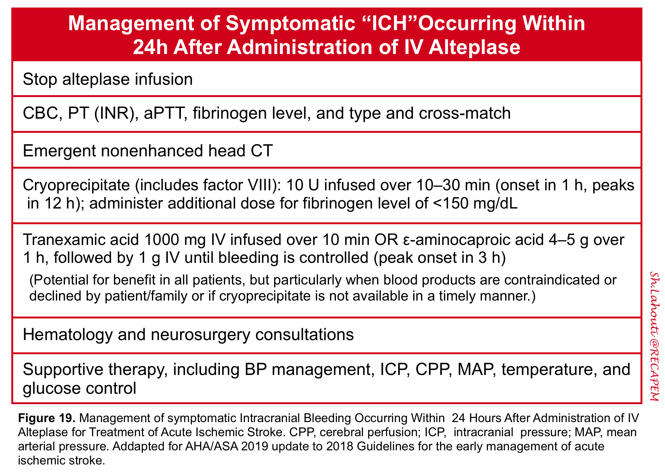
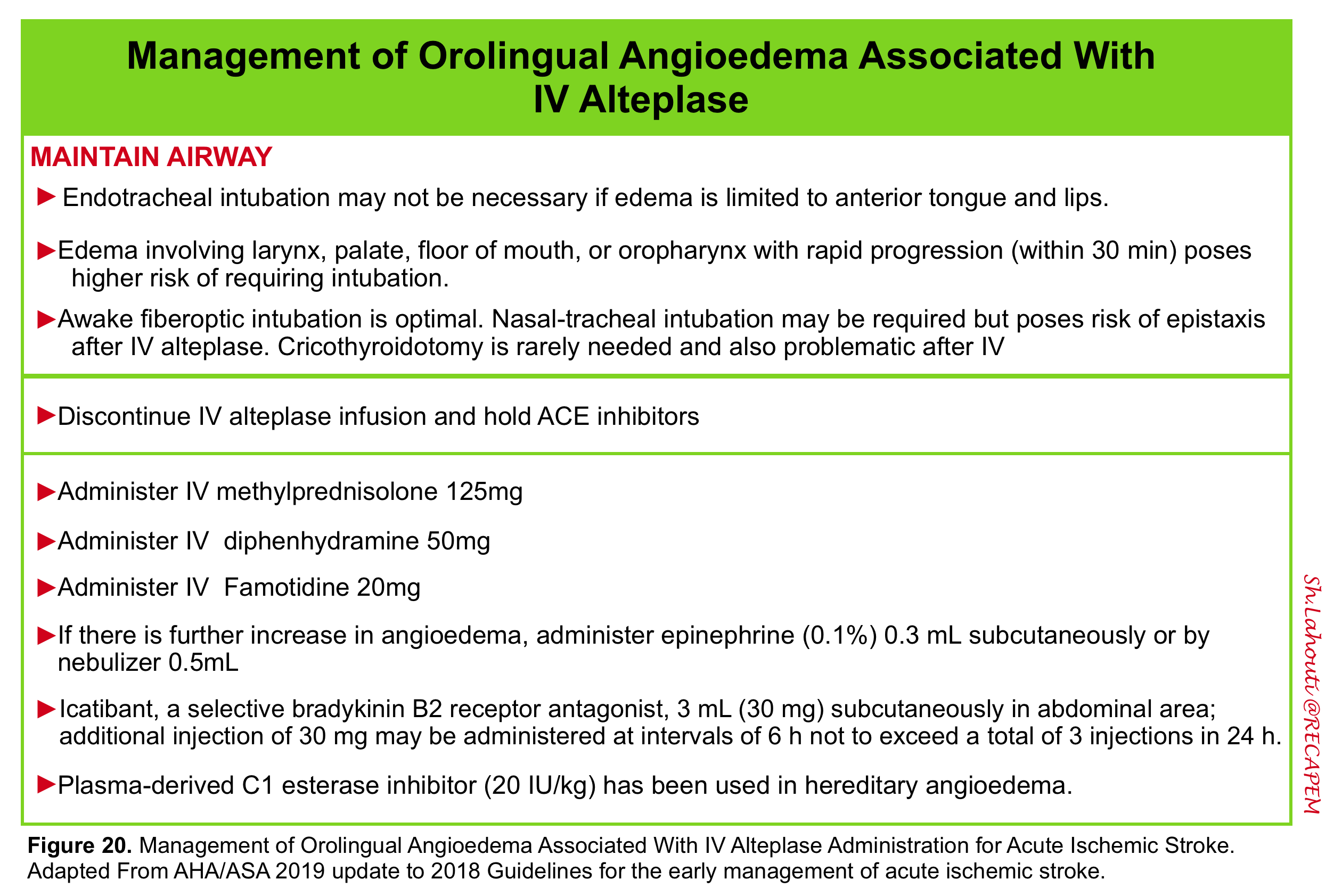
| Endovascular Thrombectomy (EVT) |
- Endovascular therapy (EVT) involves mechanical clot removal from a proximal occlusion.
- Evidence
- EVT became standard of care for LVO with the 2015 NEJM trials (≤6 h), then expanded to up to 24 h in 2018–2019 based on late-window selection by advanced imaging (DAWN, DEFUSE 3).
- Across meta-analyses, NNT ≈ 4 for significantly improved functional outcomes.
- The selection criteria included are:
- Clinical severity
- Imaging confirmation (large vessel occlusion, salvageable penumbra via advanced imaging).
- EVT became standard of care for LVO with the 2015 NEJM trials (≤6 h), then expanded to up to 24 h in 2018–2019 based on late-window selection by advanced imaging (DAWN, DEFUSE 3).
- Inclusion
- EVT is recommended for patients who meet the following core criteria. The specific time windows and imaging requirements are detailed in the table below *.
- 📍Core Patient Criteria *
- Stroke Type: Acute ischemic stroke (AIS) caused by a large vessel occlusion (LVO).
- Vessels: Proximal anterior circulation (ICA, M1 segment of MCA) or basilar artery (posterior circulation).
- Disability: NIHSS score ≥ 6 (generally; specific trials used ≥6 or ≥10 for basilar).
- Functional Status: Pre-stroke mRS score of 0-1 (independent), though EVT may be reasonable for those with mRS 2-4.
- Age: Adults (specific trials included patients up to 80+ years; pediatric recommendations exist separately).
- 🧠The Importance of the Penumbra in Ischemic Stroke Management *
- Ischemic stroke comprises core, penumbra, and normal tissue; reperfusion salvages penumbra, with benefit determined by collateral status and penumbral decay rate.
- Small core/large penumbra on advanced imaging identifies late-window candidates most likely to achieve functional recovery.
- ⏳Time-based criteria for EVT in ischemic stroke *
- 0–6 hours from LSN:
- EVT is indicated for most eligible patients with LVO, disabling symptoms, and minimal infarct core seen on non-contrast CT.
- Imaging: Plain CT to rule out hemorrhage, CTA for LVO; go directly to EVT center (do not delay for advanced imaging).
- 6–24 hours window (“extended” or “late” window) *
- EVT is considered only if advanced imaging (CT perfusion, MRI DWI/FLAIR, or multiphase CTA) shows:
- Small infarct core (e.g., <70 mL by RAPID or similar algorithms).
- Large viable penumbra (ischemic but salvageable tissue).
- Clinical–imaging mismatch (NIHSS relative to core size).
- EVT is considered only if advanced imaging (CT perfusion, MRI DWI/FLAIR, or multiphase CTA) shows:
- 0–6 hours from LSN:
- 🩻Imaging requirements for EVT in ischemic stroke *
- Non-contrast CT
- Excludes intracranial hemorrhage.
- Assesses established infarct core using ASPECTS (Alberta Stroke Program Early CT Score; ≥6 preferred for benefit).
- CT Angiogram (CTA)
- Confirms presence and site of LVO.
- Assesses for tandem lesions (extracranial/intracranial) and dissection.
- Advanced imaging for late window
- CT perfusion or MRI DWI/FLAIR *
- Measures infarct core and penumbra (thresholds: DAWN used <21 mL for >80 years and high NIHSS, <31 mL for 60–79, <51 mL for <60).
- Clinical–core mismatch: e.g., NIHSS ≥10, core <31 mL.
- Multiphase CTA:
- Evaluates collateral flow to predict tissue at risk and possible benefit.
- CT perfusion or MRI DWI/FLAIR *
- Non-contrast CT
- 📍Core Patient Criteria *
- EVT is recommended for patients who meet the following core criteria. The specific time windows and imaging requirements are detailed in the table below *.
- 🚫Relative or exclusion criteria for endovascular therapy *
- Large established infarct (ASPECTS ≤5, core >70 mL)
- Associated with a higher risk of hemorrhagic transformation and poorer outcome; EVT is generally not recommended unless compelling clinical rationale.
- Minor, nondisabling symptoms
- Benefit–risk does not favor EVT.
- Medium or distal vessel occlusions (nondominant M2, M3, A2, A3, P2, P3 segments) based on the ESCAPE-MeVO and DISTAL trials.
- Benefit–risk does not favor EVT.
- Premorbid disability (mRS >2)
- Usually excluded, but individualized if a new deficit prevents return to meaningful baseline function.
- Poor vascular access or life expectancy < 6 months.
- Other severe comorbidities limit the benefit from intervention.
- Large established infarct (ASPECTS ≤5, core >70 mL)
| Time Window | ASPECTS / Imaging Core | Additional Patient Criteria | Recommendation (COR/LOE) | Key Trial / Notes |
|---|---|---|---|---|
| 0 – 6 hours | ASPECTS 3 – 10 NCCT or MRI (no advanced imaging required) |
NIHSS ≥6, prestroke mRS 0–1, ICA or M1 occlusion | Class 1, LOE A Recommended |
HERMES, MR CLEAN, ESCAPE, SWIFT PRIME, REVASCAT |
| 0 – 6 hours (selected) | ASPECTS 0 – 2 (large core) NCCT or DWI-MRI |
Age <80 yrs, NIHSS ≥6, prestroke mRS 0–1, no significant mass effect on imaging | Class 2a, LOE B-R Reasonable |
LASTE, SELECT2, ANGEL-ASPECT (limited data; benefit magnitude smaller) |
| 0 – 6 hours | ASPECTS ≥6 | Prestroke mRS = 2, NIHSS ≥6, ICA/M1 occlusion | Class 2a, LOE B-NR Reasonable to reduce accumulated disability |
HERMES substudy, meta-analyses |
| 0 – 6 hours | ASPECTS ≥6 | Prestroke mRS 3 – 4, NIHSS ≥6, ICA/M1 occlusion | Class 2b, LOE B-NR Might be reasonable |
Retrospective/registry data; 20-30% return to premorbid mRS |
| 6 – 24 hours | ASPECTS ≥6 Requires advanced imaging: CTP or MR DWI/PWI mismatch (DAWN/DEFUSE-3 criteria) |
NIHSS ≥6, prestroke mRS 0–1, ICA/M1 occlusion, salvageable tissue | Class 1, LOE A Recommended |
DAWN, DEFUSE-3, AURORA meta-analysis |
| 6 – 24 hours (selected) | ASPECTS 3 – 5 (large core) CTP or MR DWI/PWI mismatch, or core volume criteria |
Age <80 yrs, NIHSS ≥6, prestroke mRS 0–1, no significant mass effect | Class 1, LOE A Recommended |
ANGEL-ASPECT, SELECT2 (extended window large-core trials) |
| 0 – 24 hours (basilar artery) | PC-ASPECTS ≥6 (mild-moderate ischemic damage) CTA/MRA confirms BA occlusion |
NIHSS ≥10, prestroke mRS 0–1, within 24h of onset | Class 1, LOE A Recommended |
ATTENTION, BAOCHE; NIHSS 6-9 not well established (Class 2b) |
| 0 – 6 hours (M2 branch) | ASPECTS ≥6 | Dominant M2 segment occlusion (supplying ≥50% of MCA territory), NIHSS ≥6 | Class 2a, LOE B-NR Reasonable, benefits uncertain |
HERMES pooled analysis, MR CLEAN registry |
| Any time window | Nondominant/codominant M2, distal MCA (M3), ACA (A2/A3), PCA (P2/P3) | Medium or distal vessel occlusion, often lower NIHSS | Class 3: No Benefit, LOE A Not recommended |
ESCAPE-MeVO, DISTAL trials (no benefit, potential harm with stent retrievers) |
🧠 Imaging key points:
• Early window (0-6h): NCCT or MRI sufficient; advanced imaging (CTP, MR DWI/PWI) NOT required.
• Late window (6-24h): Advanced imaging required (perfusion-core mismatch or clinical-core mismatch) to identify salvageable tissue.
• Large core (ASPECTS 0-5): EVT provides benefit but absolute rates of functional independence lower than in smaller cores; shared decision-making advised.
✅ Abbreviations: EVT = endovascular thrombectomy; ASPECTS = Alberta Stroke Program Early CT Score; PC-ASPECTS = posterior circulation ASPECTS; CTP = CT perfusion; mRS = modified Rankin Scale.
⚠️ Note: For patients with prestroke mRS 3-4 or low NIHSS, individualized decision-making is essential. The table reflects the strongest evidence from randomized trials.
Specific Indications for Posterior Circulation (Basilar Artery)
- Recommendation
- EVT is recommended (Class 1, LOE A) for patients with basilar artery occlusion presenting within 24 hours of symptom onset *.
- Specific Criteria *
- Pre-stroke mRS 0 – 1
- NIHSS score ≥ 10 at presentation
- PC-ASPECTS (posterior circulation ASPECTS) ≥ 6 (mild ischemic damage)
- Note: For patients with NIHSS 6 – 9, the effectiveness is “not well established” *.
Other Scenarios (Selected Patients)
- The 2026 guideline also provides guidance on EVT for the following situations *:
- Tandem occlusions (carotid + intracranial)
- May require acute stenting + thrombectomy; requires coordination with the neurointerventional team.
- Stroke on anticoagulation
- Relative contraindication; consult the stroke team for individualized risk assessment.
- Rapid clinical improvement
- Not typically offered EVT unless there is residual significant LVO and imaging suggests salvageable tissue.
- Tandem occlusions (carotid + intracranial)
| Clinical Scenario | Key Eligibility / Imaging | Recommendation (COR/LOE) | Supporting Evidence / Notes |
|---|---|---|---|
| Prestroke mRS = 2 (mild disability, independent but some impairment) |
Anterior circulation LVO (ICA/M1), NIHSS ≥6, ASPECTS ≥6, within 6 hours of onset | Class 2a, LOE B-NR Reasonable to reduce accumulated disability |
HERMES meta-analysis (n=199 with mRS 1-2); EVT improved outcomes vs medical therapy despite marginally worse outcomes than mRS 0 |
| Prestroke mRS = 3 – 4 (moderate to moderately severe disability; requires some assistance) |
Anterior circulation LVO, NIHSS ≥6, ASPECTS ≥6, within 6 hours (limited data for late window) | Class 2b, LOE B-NR Might be reasonable to improve functional outcomes / return to premorbid status |
Observational cohorts, Czech registry, retrospective studies: ~20-30% return to premorbid mRS; no RCTs available. Shared decision-making essential. |
| Dominant proximal M2 occlusion (supplies ≥50% of MCA territory) |
Within 6 hours, NIHSS ≥6, ASPECTS ≥6, prestroke mRS 0–1 | Class 2a, LOE B-NR Reasonable, but benefits uncertain |
HERMES pooled analysis (adjusted OR 2.39 for mRS 0-2), MR CLEAN registry; effect more pronounced in dominant M2 vs nondominant. |
| Nondominant/codominant M2, distal MCA (M3), ACA (A2/A3), PCA (P2/P3) (medium/distal vessel occlusions) |
Any time window, often lower NIHSS (median 6-7) | Class 3: No Benefit, LOE A Not recommended |
ESCAPE-MeVO and DISTAL trials: no improvement in 90-day mRS 0-1; potential harm with stent retrievers (higher mortality in ESCAPE-MeVO). |
| Basilar artery occlusion NIHSS score 6 – 9 at presentation |
PC-ASPECTS ≥6, within 24 hours, prestroke mRS 0–1 | Class 2b, LOE B-R Effectiveness not well established (uncertain) |
BAOCHE trial enrolled only 17 patients with NIHSS 6-9; limited data. For NIHSS ≥10 → Class 1 recommendation. |
| Pediatric AIS, age ≥6 years with LVO and disabling deficit |
Within 6 hours of symptom onset; PedNIHSS ≥6; imaging confirms LVO | Class 2a, LOE C-LD Can be effective to improve functional outcomes (if performed by experienced neurointerventionalists) |
Save ChildS Pro registry, retrospective multicenter studies; no RCTs. Vessel caliber near adult size after age 6. |
| Pediatric AIS (≥6 years), 6–24 hour window with salvageable brain tissue |
Perfusion imaging or DWI-FLAIR mismatch, LVO, disabling deficit | Class 2a, LOE C-LD Reasonable to improve functional outcomes |
Save ChildS Pro registry, matched case-control studies; pediatric-specific perfusion thresholds not yet established. |
| Pediatric AIS, age 28 days – 6 years with LVO and first-time seizure or disabling deficit |
Within 24 hours, salvageable brain tissue on imaging, performed by neurointerventionalist with pediatric experience | Class 2b, LOE C-LD May be reasonable (very limited evidence) |
Save ChildS Pro registry, case series; smaller vessel caliber requires high expertise. Neonates (<28 days): only case reports, no systematic evidence. |
| Large ischemic core (ASPECTS 0-5) but with severe hypodensity (≤26 HU on NCCT) |
SELECT2 exploratory analysis: CT hypodensity volume ≥26 mL at ≤26 Hounsfield units | Class 2b (caution) Diminished or no benefit; may increase cerebral edema risk |
Hypodensity threshold suggests irreversible injury; clinical judgment advised; shared decision-making. |
🧠 Key considerations for “other scenarios”:
• Prestroke disability (mRS 2-4): EVT may still restore patients to their previous functional level. Discuss goals with patient/family.
• M2 occlusions: Only dominant M2 branches show probable benefit; non-dominant M2 and distal vessels do not benefit from EVT per recent RCTs.
• Basilar artery with NIHSS 6-9: Data insufficient; decision should be individualized, considering core size and clinical trajectory.
• Pediatric EVT: Strongly recommend involvement of specialized pediatric neurointerventional teams; age <6 years carries higher technical challenges.
✅ Abbreviations: mRS = modified Rankin Scale; MCA = middle cerebral artery; ACA = anterior cerebral artery; PCA = posterior cerebral artery; PedNIHSS = Pediatric NIHSS; HU = Hounsfield units.
⚠️ Note: For prestroke mRS 3-4 and low NIHSS basilar occlusion, shared decision-making is strongly recommended. The Class 2b recommendations reflect uncertainty and require individualized risk-benefit assessment.
💡Bottom Line
- EVT is indicated for eligible patients with a disabling LVO stroke (anterior circulation or basilar artery) within 24 hours of onset.
- The 2026 guideline expands access to patients with larger cores (ASPECTS 0-5) and strongly recommends EVT for basilar artery occlusion within 24 hours *.
Complications following EVT
- Access site complications
- Groin hematoma and bleeding from the femoral access site:
- Manual pressure.
- Ultrasound to evaluate for pseudoaneurysm.
- Retroperitoneal hematoma:
- Manual pressure.
- Transfusion support.
- Check coagulation parameters (especially s/p thrombolytic administration).
- STAT CT with CT angiography.
- Possible endovascular or surgical repair.
- Femoral artery pseudoaneurysm:
- Ultrasound-guided compression.
- Thrombin injection.
- Possible open surgical repair.
- Femoral artery dissection:
- Usually observation.
- Rarely, stenting is required.
- Femoral artery occlusion:
- Evaluation by vascular surgery or interventional radiology.
- Either endovascular or surgical revascularization.
- Distal hand ischemia (s/p radial access):
- Vascular surgical consultation *.
- Groin hematoma and bleeding from the femoral access site:
- Cervical Artery Dissection
- Usually treated with observation and antiplatelet therapy.
- Rarely: carotid or vertebral stenting.
- Vasospasm
- Usually observation.
- Intra-arterial vasodilators (verapamil, nicardipine, milrinone, nimodipine) are often used.
- Vessel perforation
- Intra-procedural interventions (e.g., occlusion).
- Post-procedure CT scan.
- New embolic stroke (distal or to a new territory)
- Intra-procedural interventions (e.g., potential thrombectomy, intra-arterial thrombolytic).
- BP management *.
| Bridge Endovascular Therapy |
◾️When are both IV thrombolysis and EVT indicated?
- For eligible patients with AIS due to large vessel occlusion (LVO), the guideline recommends both IVT AND EVT– this is called “bridging therapy.”
- However, the 2026 guideline does not recommend withholding IVT to facilitate faster EVT. The combination is safe, effective, and remains the standard of care *.
◾️Indications for Bridging Therapy (Both IVT + EVT) *
- A patient should receive both IVT and EVT when they meet all of the following criteria:
- 1. IVT Eligibility Criteria (Standard)
- Time window: Within 4.5 hours of symptom onset or last known well
- Disabling neurological deficits (functional impairment)
- No intracranial hemorrhage on NCCT or MRI
- No absolute contraindications (see earlier table)
- 2. EVT Eligibility Criteria (Standard)
- Large vessel occlusion (LVO) on vascular imaging (CTA or MRA):
- Anterior circulation: ICA terminus or M1 segment of MCA
- Or basilar artery (posterior circulation)
- NIHSS score ≥ 6 (generally)
- Prestroke mRS 0–1 (independent)
- ASPECTS ≥ 3 (or selected large-core patients)
- Large vessel occlusion (LVO) on vascular imaging (CTA or MRA):
- 3. Timing
- Patient presents within 4.5 hours of onset
- EVT can be initiated immediately after IVT (no observation period)
- 1. IVT Eligibility Criteria (Standard)
| Clinical Scenario / Criteria | Key Requirements | Recommendation (COR/LOE) | Evidence / Notes |
|---|---|---|---|
| ✅ Standard Bridging (IVT + EVT) Patient eligible for both therapies |
• AIS due to LVO (ICA/M1 or basilar artery) • Within 4.5 hours of symptom onset • Disabling deficits • No IVT contraindications • NIHSS ≥6, prestroke mRS 0–1, ASPECTS ≥3 |
Class 1, LOE A Recommended |
IVT safe + improves reperfusion efficacy; HERMES, IRIS meta-analysis (bridging superior to EVT alone when IVT given early) |
| ⏱️ After IVT administration No observation period |
Do NOT wait to assess clinical response to IVT Proceed directly to EVT as rapidly as possible |
Class 1, LOE A Recommended |
Time to arterial puncture is directly associated with outcomes; any delay reduces benefit (REVASCAT, HERMES) |
| ⚠️ IVT contraindicated But EVT eligible |
LVO within 0–24 hours IVT not given due to absolute contraindication (e.g., recent surgery, coagulopathy, DOAC <48h with high risk) |
Class 2a / 1 EVT alone recommended |
EVT alone is effective even without bridging; many RCTs included IVT-ineligible patients |
| 🕒 Extended window (4.5 – 24h) IVT + EVT bridging |
• LVO + salvageable penumbra on advanced imaging (CTP or MR DWI/PWI mismatch) • IVT given per extended window criteria (TRACE-III, EXTEND) |
Class 2a / 2b, LOE B-R May be reasonable, but uncertain if EVT rapidly available |
TIMELESS trial: tenecteplase + EVT showed no benefit over placebo + EVT when rapid EVT performed. Benefit may exist when EVT delayed. |
| 💊 Choice of thrombolytic for bridging | Either alteplase (0.9 mg/kg) or tenecteplase (0.25 mg/kg) can be used before EVT | Class 1, LOE A Both recommended |
EXTEND-IA TNK, ACT trials: no difference in recanalization rates or functional outcomes between tenecteplase and alteplase before EVT |
| 📉 Mild deficits (NIHSS <6) with LVO | Disabling deficit assessment is key; if deficits are NOT disabling → IVT not recommended (Class 3). If disabling despite low NIHSS → bridging may be considered. | Class 2b Individualized decision |
Limited data; TEMPO-2 (minor stroke with occlusion) did not show benefit of tenecteplase over standard care |
| 🧠 Large ischemic core (ASPECTS 0–5) within 4.5 hours |
Patients with large core who are IVT-eligible and have LVO may receive bridging, but benefit magnitude smaller | Class 2a, LOE B-R Reasonable |
SELECT2, ANGEL-ASPECT, LASTE included IVT-eligible patients; absolute functional independence rates lower than small-core trials |
| 🧠 Basilar artery occlusion within 4.5 hours |
IVT (if eligible) + EVT (up to 24h) – bridging recommended when patient presents early | Class 1, LOE A Recommended for NIHSS ≥10 |
ATTENTION, BAOCHE; IVT was used in a proportion of patients. No specific RCT of bridging vs EVT alone in posterior circulation. |
🔑 Key Takeaway from the 2026 Guideline (Section 4.7.1):
• Do NOT skip IVT to save time. Bridging therapy (IVT + EVT) is superior to EVT alone when IVT can be given within the 4.5-hour window.
• Do NOT delay EVT to observe clinical response after IVT. Time is brain — proceed directly to thrombectomy.
• For extended window (4.5–24h) with advanced imaging selection, the benefit of adding IVT to EVT is less certain, especially if EVT is rapidly available (TIMELESS trial).
• Both alteplase and tenecteplase (0.25 mg/kg) are acceptable bridging agents.
✅ Abbreviations: IVT = intravenous thrombolysis; EVT = endovascular thrombectomy; LVO = large vessel occlusion; mRS = modified Rankin Scale; ASPECTS = Alberta Stroke Program Early CT Score.
⚠️ Important: Bridging therapy is indicated for eligible patients presenting within 4.5 hours. For patients presenting beyond 4.5 hours up to 24 hours, EVT alone is standard; IVT may be considered in select patients with advanced imaging mismatch (see extended window recommendations).
⭐️ Important Practical Points
- Do not delay EVT to “watch” if the patient improves after IVT.
- IVT should be administered as rapidly as possible, without observation to assess clinical response or delay in initiating EVT (if eligible) *.
- Safety
- Bridging therapy does not increase the risk of symptomatic intracranial hemorrhage (sICH) compared to EVT alone, based on the meta-analysis of 6 RCTs *.
- Extended window (4.5 – 24 hours)
- For patients who present in the extended window and receive IVT based on advanced imaging (e.g., TRACE-III, TIMELESS), bridging therapy may still be used, but the benefit is less certain if rapid EVT is performed (TIMELESS trial showed no benefit of tenecteplase over placebo when EVT was rapidly available) *.
Other therapeutic measures
◾️Antiplatlet Therapy after IVT (see the following table)
🧠 Post-EVT Antithrombotic Strategy 2026 AHA/ASA Guideline
◾️Antithrombotic medications after EVT (see the following table)
💊 Post-Thrombolysis (Alteplase / Tenecteplase) Antithrombotic Strategy 2026 AHA/ASA Update
- Hyperthermia (temperature > 38 °C) contributes to neuronal injury 6 in patients with acute stroke via several different mechanisms (more on this here).
- Elevated temperature (> 38 °C) should be evaluated and treated aggressively.
- Do NOT use induced hypothermia or prophylactic fever prevention in normothermic patients – no benefit and potential harm *.
- Treat hypoglycemia (<60 mg/dL) immediately *.
- For hyperglycemia, target glucose 140-180 mg/dL*.
- Intensive glucose control to 80-130 mg/dL is NOT recommended – it does not improve outcomes and increases risk of severe hypoglycemia *.
- Patients treated with thrombolysis or successful endovascular thrombectomy: delay chemical DVT prophylaxis for 24 hours *.
- Patients unable to receive chemical DVT prophylaxis should be managed with intermittent pneumatic compression.
- Low-molecular-weight heparin (e.g., enoxaparin 40mg daily) is preferred for DVT prophylaxis *.
- Treat unprovoked seizures after AIS with antiseizure medication (individualize choice and duration) *.
- Do NOT use prophylactic antiseizure medication routinely – it does not prevent seizures or improve outcomes.
- NOTE: Seizure at onset is NOT a contraindication to IVT if deficits are due to stroke rather than postictal state *.
Nondisabling AIS and TIAs
Up to half of all ischemic strokes are non-disabling *. Although clinical deficits are mild, the 30-day risk of neurologic deterioration or disabling stroke is about 4-5% *. ED priorities include precise phenotyping, urgent vascular imaging when indicated, early secondary prevention, and reliable short-interval follow-up.
- TIA and nondisabling stroke live on the same continuum.
- Non-disabling strokes and TIAs share pathophysiology, prognosis, and treatments.
- The DOUBT study found that 13% of TIA presentations as short as 5 minutes in duration are subsequently found to have evidence of stroke lesions on MRI. 2-17% of patients with TIA or minor stroke suffer a subsequent disabling stroke within 90 days, most within 48 hours *.
- Non-disabling strokes and TIAs share pathophysiology, prognosis, and treatments.
- The 2026 guideline recommends against IV thrombolysis for non-disabling deficits (Class 3) and instead emphasizes urgent evaluation and DAPT for secondary prevention *.
◾️Pathophysiology
- As already discussed, the pathophysiology of the “AIS” and “ TIA” can be classified into (TOAST classification system 37):
- Large artery atherosclerosis
- Cardioembolism
- Small-vessel occlusion
- Stroke of another determined etiology
- Stroke of undetermined etiology
- Two or more causes identified
- Negative evaluation
- Incomplete evaluation
- Sometimes, despite a thorough evaluation, the etiology of ischemic stroke is not well-defined; hence called cryptogenic stroke.
- It is defined as a brain infarction that is not attributable to a source of definite cardioembolism, large artery atherosclerosis, or small artery disease despite a thorough vascular, cardiac, and serologic evaluation 38.
- The pathophysiology of cryptogenic stroke is likely heterogeneous.
- Proposed mechanisms include cardiac embolism secondary to occult paroxysmal atrial fibrillation, aortic atheromatous disease, or other cardiac sources, paradoxical embolism from atrial septal abnormalities such as patent foramen ovale, hypercoagulable states, and preclinical or subclinical cerebrovascular disease.
- It has been shown that a significant proportion of cryptogenic strokes adhere to embolic infarct topography on brain imaging.
- Cryptogenic stroke is a diagnosis of exclusion.
- The acute management of cryptogenic stroke is similar to that of other ischemic stroke subtypes.
Approach to diagnosis and management
Diagnosis and initial management
- When suspected, perform STAT NCCT to rule out ICH and other etiologies.
- Why Persistent Deficits and High-Risk TIA Features Matter
- The Common Message
- Persistent non-disabling deficits – even when “mild” – raise the pretest probability of a treatable lesion (e.g., symptomatic carotid stenosis, intracranial atherosclerosis, or cervical artery dissection), creating an opportunity for secondary prevention *.
- Resolved high-risk features – true motor weakness (often described as heaviness), speech or language disturbance, major visual field loss, or depressed consciousness – suggest a vulnerable vascular territory at high risk for subsequent stroke from ongoing pathology *.
- The Common Message
- Urgent Actions Required
- The Goal
- Prevent the next stroke event – the highest risk period is within the first 48 hours to one week.
- Why Persistent Deficits and High-Risk TIA Features Matter
⚠️High-risk TIAs
- It has been shown that the most widely used prediction tool for risk stratification (ABCD2 score) does not provide an accurate estimate of stroke risk, and that clinical decisions based on an ABCD2 score cut-off are subject to significant misclassification error. Clinical judgment, along with the integration of certain clinical and imaging features, is more accurate for predicting the risk of cerebrovascular events 40.
| Category | High-Risk Feature | Clinical Implication |
|---|---|---|
| Clinical Features Urgent |
• Motor weakness (hemiparesis, facial droop, arm/leg drift) • Speech disturbance (aphasia, dysarthria) • Vision loss (monocular blindness / amaurosis fugax, homonymous hemianopia) • Gait disturbance (ataxia, imbalance, inability to walk) • Crescendo TIAs (≥2 episodes within 24-48 hours, each lasting ≥10 minutes) |
Highest risk of early recurrent stroke; requires immediate evaluation and admission or same-day rapid-access clinic |
| ABCD² Score ≥4 Class 1 |
• Age ≥60 years (1 point) • Blood pressure ≥140/90 mmHg (1 point) • Clinical features: unilateral weakness (2 points) or speech impairment without weakness (1 point) • Duration ≥60 minutes (2 points) or 10-59 minutes (1 point) • Diabetes (1 point) Total score ≥4 (2-day stroke risk ~4-8%) |
Sensitivity 100% for 7-day stroke when score 0-3 (NPV 100%). However, low score does NOT rule out high-risk pathology. |
| Vascular Features Urgent |
• Symptomatic carotid stenosis ≥50% • Intracranial stenosis ≥50-70% • Large vessel occlusion (LVO) on CTA/MRA • Arterial dissection (carotid or vertebral) |
Urgent revascularization (CEA/CAS) may be indicated for ≥70% stenosis. DAPT for 90 days for intracranial stenosis. |
| Cardiac Features Urgent |
• Atrial fibrillation (paroxysmal or persistent) • Mechanical heart valve • Left ventricular thrombus • Cardioembolic source (valve, myxoma, endocarditis) |
Anticoagulation indicated (DOAC preferred for non-valvular AF). DO NOT use antiplatelet monotherapy. |
| Imaging Features Urgent |
• DWI-positive lesion on MRI (Reclassifies event as ischemic stroke, not TIA, by tissue-based definition) |
Same risk of recurrence as minor stroke. Requires aggressive secondary prevention (DAPT, statin, risk factor control). |
• The ABCD² score lacks sufficient sensitivity and specificity to reliably triage TIA patients.
• Patients with low ABCD² scores (0-3) may still harbor significant pathology (e.g., carotid stenosis, atrial fibrillation, DWI-positive lesion).
• Do NOT rely on ABCD² score alone – clinical judgment and high-risk features (motor weakness, speech disturbance, vision loss, crescendo TIAs) should guide urgency.
A TIA is considered HIGH-RISK if ANY of the following are present:
• Clinical red flags: motor weakness, speech disturbance, vision loss, gait disturbance, crescendo TIAs
• ABCD² score ≥4
• High-risk etiology: carotid stenosis ≥50%, atrial fibrillation, intracranial stenosis, LVO, dissection, DWI-positive lesion
Action: Admit or arrange same-day rapid-access TIA clinic evaluation. Start DAPT (aspirin + clopidogrel) for 21 days unless anticoagulation indicated. Urgent MRI/CTA and cardiac monitoring required.
✅ Key takeaway: High-risk TIA requires urgent evaluation (within 24-48 hours) with MRI/CTA, cardiac monitoring, and DAPT (or anticoagulation if AF). Do NOT rely on ABCD² score alone – clinical red flags (motor weakness, speech disturbance, vision loss, crescendo TIAs) and high-risk features (carotid stenosis, AF) mandate same-day evaluation regardless of score.
Evaluation
Determining the pathophysiology of ischemic stroke and TIA
- Cardiac monitoring for possible occult atrial fibrillation
- Echocardiography (TTE/TEE) to identify the possible source of cardioembolism (figure 4).
- Vascular study (Duplex ultrasound of neck, CTA or MRA of neck and head arteries) to determine the status of major cerebral vessels.
- Blood test, including hypercoagulable studies in select patients.
Treatment pathway based on stroke etiology
- Large-artery atherosclerosis (carotid/intracranial):
- Clues: prior TIA, bruits, focal cortical signs.
- Action: CTA head/neck to find carotid stenosis/dissection.
- CEA: symptomatic ICA 70–99% stenosis likely to need surgery/stenting; 50–70% often considered for surgery/stenting.
- Timing: avoid the first 48 h after stroke; target within ~2 weeks before benefit wanes.
- Antithrombotic while waiting for CEA (pre-op): single antiplatelet plus statin and risk-factor control.
- Lacunar/small-vessel:
- Clues: pure motor/sensory syndromes, small deep infarcts.
- Action: DAPT and aggressive risk-factor management.
- Cardioembolic (AF, cardiomyopathy, valve/aortic thrombus, PFO):
- Action: ECG/monitoring now; plan oral anticoagulation if indicated when safe (see below), even if imaging looks “lacunar”, may require surgical repair of structural cardiac lesions.
- Other/cryptogenic (dissection, thrombophilia, hyperviscosity; higher in young):
- Clues: neck pain/trauma for dissection; hypercoagulable risk factors; migraine-like prodromes with sudden negative deficits.
- ED actions: CTA head/neck to assess dissection/stenosis; short-course (21 days) DAPT for TIA/nondisabling stroke unless cardioembolic mechanism surfaces; coordinate targeted work-up with consultant.
◾️Sorting out the etiology will determine the line of secondary prevention and its timeline 👇.
Secondary Prevention
- 🔪Carotid endarterectomy (CEA) in nondisabling stroke / high-risk TIA
- Carotid endarterectomy remains one of the few surgical interventions in stroke care that meaningfully alters outcomes. Patients with symptomatic internal carotid artery stenosis of 70–99% derive the greatest absolute risk reduction in recurrent stroke with early CEA. Selected patients with 50–69% stenosis may also benefit, depending on plaque/composition/stability, comorbidities, and symptoms.
- 🕰️Timing is critical: CEA is typically deferred for the first 48 hours following ischemic stroke to avoid reperfusion injury in the “hot brain” phase, then performed as soon as safely feasible—ideally within 7–14 days of the index event. The benefit declines substantially with delay beyond two weeks.
- Carotid endarterectomy remains one of the few surgical interventions in stroke care that meaningfully alters outcomes. Patients with symptomatic internal carotid artery stenosis of 70–99% derive the greatest absolute risk reduction in recurrent stroke with early CEA. Selected patients with 50–69% stenosis may also benefit, depending on plaque/composition/stability, comorbidities, and symptoms.
- 💊Medical therapy for nondisabling stroke: Secondary prevention (once intracranial hemorrhage is ruled out)
- Load antiplatelets in the ED and tailor the regimen to the mechanism.
- Most patients without endarterectomy plans or atrial fibrillation benefit from a short course of dual antiplatelet therapy (DAPT)—typically 21 days—then de-escalation to monotherapy.
- Pragmatically, aspirin 160–325 mg loading then 81 mg daily combined with clopidogrel 300 mg loading then 75 mg daily is common; aspirin with ticagrelor 180 mg loading then 90 mg twice daily is a reasonable alternative. Benefit is front-loaded in the first three weeks; bleeding risk rises after, which is why DAPT is not continued indefinitely as a standard.
- Use single antiplatelet therapy if carotid surgery is anticipated, extend dual antiplatelet therapy when intracranial atherosclerosis is the culprit per local protocol, and/or transition to anticoagulation when cardioembolic sources such as atrial fibrillation are identified, and it is safe to do so.
- For symptomatic cervical artery dissection with TIA or nondisabling stroke, treatment typically involves a short course of DAPT, while asymptomatic dissection commonly receives single-agent therapy for months in coordination with the stroke team.
- Most patients without endarterectomy plans or atrial fibrillation benefit from a short course of dual antiplatelet therapy (DAPT)—typically 21 days—then de-escalation to monotherapy.
- Load antiplatelets in the ED and tailor the regimen to the mechanism.
Antiplatelet therapy for nondisabling stroke
- 💊Single antiplatelet medication options
- ASA 160–325 mg load → 81 mg daily, or
- Clopidogrel 300 mg load → 75 mg daily (use when ASA allergy/intolerance or DAPT chosen).
- 💊💊DAPT (short course ~21–28 days):
- ASA + Clopidogrel (default in many pathways), then step down to a single antiplatelet agent.
- Consider ASA + Ticagrelor, where clopidogrel resistance/genotype is a concern *, *.
- Avoid DAPT in disabling infarcts (higher bleed risk into established core) *.
- Etiology-specific nuances:
- Intracranial atherosclerosis: consider DAPT ~90 days *.
- Cervical artery dissection: often DAPT ~21 days for TIA/nondisabling stroke; some specialists prefer anticoagulation; asymptomatic dissections frequently single antiplatelet agent ≥6 months.
- Peri-carotid endarterectomy: typically single antiplatelet pre-op (confirm with vascular/stroke protocols) *.
📖An algorithm for antiplatelet therapy can be found on page 63 of the 2026 AHA/ASA guidelines here.
Anticoagulation after nondisabling stroke
- Cardioembolic sources (e.g., AF):
- Oral anticoagulation for secondary prevention; initiation/timing individualized (consider infarct size, hemorrhagic risk, and neurology guidance) but typically within 4 days; the trend as of 2025 is to start a DOAC as soon as 24 hrs after the stroke initiation for smaller infarcts with low hemorrhagic transformation risk *,*.
- The “1-2-3-4-Day” rule proposes starting DOACs after ischemic stroke with atrial fibrillation at 1, 2, 3, or 4 days post-event, depending on stroke severity (TIA, mild, moderate, or severe, respectively), and was associated with reduced recurrent stroke or systemic embolism risk without increased major bleeding in both Japanese and European cohorts *.
Admission vs. discharge
◾️🏥Admission Criteria for Non-Disabling Stroke
- Hospital admission or observation is recommended for patients with any of the following:
- Symptomatic extracranial or intracranial stenosis >50%
- Acute infarction on MRI
- New-onset atrial fibrillation or other cardioembolic source
- Recurrent (dual) TIAs or crescendo symptoms
- Severe or labile hypertension
- Metabolic derangements or medical instability
- Uncertain diagnosis or unreliable follow-up
- Institutional barriers to rapid outpatient evaluation
◾️Safe discharge criteria for nondisabling stroke (with follow-up ≤48 hours)
- Selected patients with nondisabling stroke can be discharged safely from the ED when all of the following are satisfied:
- Mild neurological deficit (NIHSS ≤5)
- Stable neurological symptoms without recurrence, progression, or fluctuation
- Brain imaging (CT/MRI) excludes hemorrhage, large vessel occlusion, or high-risk pathology (e.g., >50% carotid stenosis)
- Lab tests show no significant acute abnormalities (e.g., no atrial fibrillation, troponin elevation, or metabolic derangements)
- Initiation of secondary prevention: antiplatelet/anticoagulation and statin as indicated
- Expedited outpatient follow-up arranged within 24-72 hours with a stroke or neurology clinic
2026 AHA/ASA Guideline for AIS Management
Media
Imaging of acute ischemic stroke
Evolution from acute to chronic
Going further
- NeuroEMCrit – Time is Brain – Acute Ischemic Stroke Part I
- Paradigm Shift in Ischemic Stroke Management-Disabling Strokes (Emergency Medicine Cases)
- TIA Update (Emergency medicine case)
- ED stroke management in the age of endovascular therapy (Emergency medicine cases)
- NeuroEMCrit- Acute ischemic stroke part I
References
1. Krishnamurthi RV, Feigin VL, Forouzanfar MH, Mensah GA, Connor M, Bennett DA, Moran AE, Sacco RL, Anderson LM, Truelsen T, O’Donnell M, Venketasubramanian N, Barker-Collo S, Lawes CM, Wang W, Shinohara Y, Witt E, Ezzati M, Naghavi M, Murray C; Global Burden of Diseases, Injuries, Risk Factors Study 2010 (GBD 2010); GBD Stroke Experts Group. Global and regional burden of first-ever ischaemic and haemorrhagic stroke during 1990-2010: findings from the Global Burden of Disease Study 2010. Lancet Glob Health. 2013 Nov;1(5):e259-81. doi: 10.1016/S2214-109X(13)70089-5. Epub 2013 Oct 24. PMID: 25104492; PMCID: PMC4181351
2. Ishihara T, Sato S, Uehara T, Ohara T, Hayakawa M, Kimura K, Okada Y, Hasegawa Y, Tanahashi N, Suzuki A, Nakagawara J, Arii K, Nagahiro S, Ogasawara K, Uchiyama S, Matsumoto M, Iihara K, Toyoda K, Minematsu K; PROMISE-TIA Study Investigators. Significance of Nonfocal Symptoms in Patients With Transient Ischemic Attack. Stroke. 2018 Aug;49(8):1893-1898. doi: 10.1161/STROKEAHA.118.022009. PMID: 30012818
3. Easton JD, Saver JL, Albers GW, Alberts MJ, Chaturvedi S, Feldmann E, Hatsukami TS, Higashida RT, Johnston SC, Kidwell CS, Lutsep HL, Miller E, Sacco RL; American Heart Association; American Stroke Association Stroke Council; Council on Cardiovascular Surgery and Anesthesia; Council on Cardiovascular Radiology and Intervention; Council on Cardiovascular Nursing; Interdisciplinary Council on Peripheral Vascular Disease. Definition and evaluation of transient ischemic attack: a scientific statement for healthcare professionals from the American Heart Association/American Stroke Association Stroke Council; Council on Cardiovascular Surgery and Anesthesia; Council on Cardiovascular Radiology and Intervention; Council on Cardiovascular Nursing; and the Interdisciplinary Council on Peripheral Vascular Disease. The American Academy of Neurology affirms the value of this statement as an educational tool for neurologists. Stroke. 2009 Jun;40(6):2276-93. doi: 10.1161/STROKEAHA.108.192218. Epub 2009 May 7. PMID: 19423857
4. Sacco RL, Kasner SE, Broderick JP, Caplan LR, Connors JJ, Culebras A, Elkind MS, George MG, Hamdan AD, Higashida RT, Hoh BL, Janis LS, Kase CS, Kleindorfer DO, Lee JM, Moseley ME, Peterson ED, Turan TN, Valderrama AL, Vinters HV; American Heart Association Stroke Council, Council on Cardiovascular Surgery and Anesthesia; Council on Cardiovascular Radiology and Intervention; Council on Cardiovascular and Stroke Nursing; Council on Epidemiology and Prevention; Council on Peripheral Vascular Disease; Council on Nutrition, Physical Activity and Metabolism. An updated definition of stroke for the 21st century: a statement for healthcare professionals from the American Heart Association/American Stroke Association. Stroke. 2013 Jul;44(7):2064-89. doi: 10.1161/STR.0b013e318296aeca. Epub 2013 May 7. Erratum in: Stroke. 2019 Aug;50(8):e239. PMID: 23652265.
5. Goyal N, Tsivgoulis G, Male S, Metter EJ, Iftikhar S, Kerro A, Chang JJ, Frey JL, Triantafyllou S, Papadimitropoulos G, Abedi V, Alexandrov AW, Alexandrov AV, Zand R. FABS: An Intuitive Tool for Screening of Stroke Mimics in the Emergency Department. Stroke. 2016
6. Powers WJ, Rabinstein AA, Ackerson T, Adeoye OM, Bambakidis NC, Becker K, Biller J, Brown M, Demaerschalk BM, Hoh B, Jauch EC, Kidwell CS, Leslie-Mazwi TM, Ovbiagele B, Scott PA, Sheth KN, Southerland AM, Summers DV, Tirschwell DL. Guidelines for the Early Management of Patients With Acute Ischemic Stroke: 2019 Update to the 2018 Guidelines for the Early Management of Acute Ischemic Stroke: A Guideline for Healthcare Professionals From the American Heart Association/American Stroke Association. Stroke. 2019 Dec;50(12):e344-e418. doi: 10.1161/STR.0000000000000211. Epub 2019 Oct 30. Erratum in: Stroke. 2019 Dec;50(12):e440-e441. PMID: 31662037
7. Potter CA, Vagal AS, Goyal M, Nunez DB, Leslie-Mazwi TM, Lev MH. CT for Treatment Selection in Acute Ischemic Stroke: A Code Stroke Primer. Radiographics. 2019 Oct;39(6):1717-1738. doi: 10.1148/rg.2019190142. PMID: 31589578
8. Kamalian S, Lev MH. Stroke Imaging. Radiol Clin North Am. 2019 Jul;57(4):717-732. doi: 10.1016/j.rcl.2019.02.001. Epub 2019 Apr 8. PMID: 31076028.
9. Schellinger PD, Bryan RN, Caplan LR, Detre JA, Edelman RR, Jaigobin C, Kidwell CS, Mohr JP, Sloan M, Sorensen AG, Warach S; Therapeutics and Technology Assessment Subcommittee of the American Academy of Neurology. Evidence-based guideline: The role of diffusion and perfusion MRI for the diagnosis of acute ischemic stroke: report of the Therapeutics and Technology Assessment Subcommittee of the American Academy of Neurology. Neurology. 2010 Jul 13;75(2):177-85. doi: 10.1212/WNL.0b013e3181e7c9dd. Erratum in: Neurology. 2010 Sep 7;75(10):938. PMID: 20625171; PMCID: PMC2905927
10. Wen HM, Lam WW, Rainer T, Fan YH, Leung TW, Chan YL, Wong KS. Multiple acute cerebral infarcts on diffusion-weighted imaging and risk of recurrent stroke. Neurology. 2004 Oct 12;63(7):1317-9. doi: 10.1212/01.wnl.0000140490.22251.b6. PMID: 15477564.
11. Sylaja PN, Coutts SB, Subramaniam S, Hill MD, Eliasziw M, Demchuk AM; VISION Study Group. Acute ischemic lesions of varying ages predict risk of ischemic events in stroke/TIA patients. Neurology. 2007 Feb 6;68(6):415-9. doi: 10.1212/01.wnl.0000252938.76188.52. PMID: 17283315
12. Thomalla G, Fiebach JB, Østergaard L, Pedraza S, Thijs V, Nighoghossian N, Roy P, Muir KW, Ebinger M, Cheng B, Galinovic I, Cho TH, Puig J, Boutitie F, Simonsen CZ, Endres M, Fiehler J, Gerloff C; WAKE-UP investigators. A multicenter, randomized, double-blind, placebo-controlled trial to test efficacy and safety of magnetic resonance imaging-based thrombolysis in wake-up stroke (WAKE-UP). Int J Stroke. 2014 Aug;9(6):829-36. doi: 10.1111/ijs.12011. Epub 2013 Mar 12. PMID: 23490032
13. Saver JL, Fonarow GC, Smith EE, Reeves MJ, Grau-Sepulveda MV, Pan W, Olson DM, Hernandez AF, Peterson ED, Schwamm LH. Time to treatment with intravenous tissue plasminogen activator and outcome from acute ischemic stroke. JAMA. 2013 Jun 19;309(23):2480-8. doi: 10.1001/jama.2013.6959. PMID: 23780461
14. Emberson J, Lees KR, Lyden P, Blackwell L, Albers G, Bluhmki E, Brott T, Cohen G, Davis S, Donnan G, Grotta J, Howard G, Kaste M, Koga M, von Kummer R, Lansberg M, Lindley RI, Murray G, Olivot JM, Parsons M, Tilley B, Toni D, Toyoda K, Wahlgren N, Wardlaw J, Whiteley W, del Zoppo GJ, Baigent C, Sandercock P, Hacke W; Stroke Thrombolysis Trialists’ Collaborative Group. Effect of treatment delay, age, and stroke severity on the effects of intravenous thrombolysis with alteplase for acute ischaemic stroke: a meta-analysis of individual patient data from randomised trials. Lancet. 2014 Nov 29;384(9958):1929-35. doi: 10.1016/S0140-6736(14)60584-5. Epub 2014 Aug 5. PMID: 25106063; PMCID: PMC4441266
15. National Institute of Neurological Disorders and Stroke rt-PA Stroke Study Group. Tissue plasminogen activator for acute ischemic stroke. N Engl J Med. 1995 Dec 14;333(24):1581-7. doi: 10.1056/NEJM199512143332401. PMID: 7477192
16. Mazya M, Egido JA, Ford GA, Lees KR, Mikulik R, Toni D, Wahlgren N, Ahmed N; SITS Investigators. Predicting the risk of symptomatic intracerebral hemorrhage in ischemic stroke treated with intravenous alteplase: safe Implementation of Treatments in Stroke (SITS) symptomatic intracerebral hemorrhage risk score. Stroke. 2012 Jun;43(6):1524-31. doi: 10.1161/STROKEAHA.111.644815. Epub 2012 Mar 22. Erratum in: Stroke. 2012 Sep;43(9):e102. PMID: 22442178
17. Mazya M, Egido JA, Ford GA, Lees KR, Mikulik R, Toni D, Wahlgren N, Ahmed N; SITS Investigators. Predicting the risk of symptomatic intracerebral hemorrhage in ischemic stroke treated with intravenous alteplase: safe Implementation of Treatments in Stroke (SITS) symptomatic intracerebral hemorrhage risk score. Stroke. 2012 Jun;43(6):1524-31. doi: 10.1161/STROKEAHA.111.644815. Epub 2012 Mar 22. Erratum in: Stroke. 2012 Sep;43(9):e102. PMID: 22442178
18. Seners P, Turc G, Maïer B, Mas JL, Oppenheim C, Baron JC. Incidence and Predictors of Early Recanalization After Intravenous Thrombolysis: A Systematic Review and Meta-Analysis. Stroke. 2016 Sep;47(9):2409-12. doi: 10.1161/STROKEAHA.116.014181. Epub 2016 Jul 26. PMID: 27462117
19. Molina CA, Montaner J, Arenillas JF, Ribo M, Rubiera M, Alvarez-Sabín J. Differential pattern of tissue plasminogen activator-induced proximal middle cerebral artery recanalization among stroke subtypes. Stroke. 2004 Feb;35(2):486-90. doi: 10.1161/01.STR.0000110219.67054.BF. Epub 2004 Jan 5. PMID: 14707233
20. Overgaard K. Thrombolytic therapy in experimental embolic stroke. Cerebrovasc Brain Metab Rev. 1994 Fall;6(3):257-86. PMID: 7811566.
21. Whiteley WN, Emberson J, Lees KR, Blackwell L, Albers G, Bluhmki E, Brott T, Cohen G, Davis S, Donnan G, Grotta J, Howard G, Kaste M, Koga M, von Kummer R, Lansberg MG, Lindley RI, Lyden P, Olivot JM, Parsons M, Toni D, Toyoda K, Wahlgren N, Wardlaw J, Del Zoppo GJ, Sandercock P, Hacke W, Baigent C; Stroke Thrombolysis Trialists’ Collaboration. Risk of intracerebral haemorrhage with alteplase after acute ischaemic stroke: a secondary analysis of an individual patient data meta-analysis. Lancet Neurol. 2016 Aug;15(9):925-933. doi: 10.1016/S1474-4422(16)30076-X. Epub 2016 Jun 8. PMID: 27289487
22. Whiteley WN, Slot KB, Fernandes P, Sandercock P, Wardlaw J. Risk factors for intracranial hemorrhage in acute ischemic stroke patients treated with recombinant tissue plasminogen activator: a systematic review and meta-analysis of 55 studies. Stroke. 2012 Nov;43(11):2904-9. doi: 10.1161/STROKEAHA.112.665331. Epub 2012 Sep 20. PMID: 22996959
23. Prabhakaran S, Ruff I, Bernstein RA. Acute stroke intervention: a systematic review. JAMA. 2015 Apr 14;313(14):1451-62. doi: 10.1001/jama.2015.3058. PMID: 25871671
24. Re-examining Acute Eligibility for Thrombolysis (TREAT) Task Force:, Levine SR, Khatri P, Broderick JP, Grotta JC, Kasner SE, Kim D, Meyer BC, Panagos P, Romano J, Scott P; NINDS rt-PA Stroke Trial Investigators. Review, historical context, and clarifications of the NINDS rt-PA stroke trials exclusion criteria: Part 1: rapidly improving stroke symptoms. Stroke. 2013 Sep;44(9):2500-5. doi: 10.1161/STROKEAHA.113.000878. Epub 2013 Jul 11. Erratum in: Stroke. 2014 Mar;45(3):e52. PMID: 23847249; PMCID: PMC3789593
25. Whiteley WN, Emberson J, Lees KR, Blackwell L, Albers G, Bluhmki E, Brott T, Cohen G, Davis S, Donnan G, Grotta J, Howard G, Kaste M, Koga M, von Kummer R, Lansberg MG, Lindley RI, Lyden P, Olivot JM, Parsons M, Toni D, Toyoda K, Wahlgren N, Wardlaw J, Del Zoppo GJ, Sandercock P, Hacke W, Baigent C; Stroke Thrombolysis Trialists’ Collaboration. Risk of intracerebral haemorrhage with alteplase after acute ischaemic stroke: a secondary analysis of an individual patient data meta-analysis. Lancet Neurol. 2016 Aug;15(9):925-933. doi: 10.1016/S1474-4422(16)30076-X. Epub 2016 Jun 8. PMID: 27289487
26. Khatri P, Kleindorfer DO, Devlin T, Sawyer RN Jr, Starr M, Mejilla J, Broderick J, Chatterjee A, Jauch EC, Levine SR, Romano JG, Saver JL, Vagal A, Purdon B, Devenport J, Pavlov A, Yeatts SD; PRISMS Investigators. Effect of Alteplase vs Aspirin on Functional Outcome for Patients With Acute Ischemic Stroke and Minor Nondisabling Neurological Deficits: The PRISMS Randomized Clinical Trial. JAMA. 2018 Jul 10;320(2):156-166. doi: 10.1001/jama.2018.8496. PMID: 29998337; PMCID: PMC6583516
27. Thomalla G, Simonsen CZ, Boutitie F, Andersen G, Berthezene Y, Cheng B, Cheripelli B, Cho TH, Fazekas F, Fiehler J, Ford I, Galinovic I, Gellissen S, Golsari A, Gregori J, Günther M, Guibernau J, Häusler KG, Hennerici M, Kemmling A, Marstrand J, Modrau B, Neeb L, Perez de la Ossa N, Puig J, Ringleb P, Roy P, Scheel E, Schonewille W, Serena J, Sunaert S, Villringer K, Wouters A, Thijs V, Ebinger M, Endres M, Fiebach JB, Lemmens R, Muir KW, Nighoghossian N, Pedraza S, Gerloff C; WAKE-UP Investigators. MRI-Guided Thrombolysis for Stroke with Unknown Time of Onset. N Engl J Med. 2018 Aug 16;379(7):611-622. doi: 10.1056/NEJMoa1804355. Epub 2018 May 16. PMID: 29766770
28. Marshall RS. Image-Guided Intravenous Alteplase for Stroke – Shattering a Time Window. N Engl J Med. 2019 May 9;380(19):1865-1866. doi: 10.1056/NEJMe1904791. PMID: 31067378
29. Engelter ST, Dallongeville J, Kloss M, Metso TM, Leys D, Brandt T, Samson Y, Caso V, Pezzini A, Sessa M, Beretta S, Debette S, Grond-Ginsbach C, Metso AJ, Thijs V, Lamy C, Medeiros E, Martin JJ, Bersano A, Tatlisumak T, Touzé E, Lyrer PA; Cervical Artery Dissection and Ischaemic Stroke Patients-Study Group. Thrombolysis in cervical artery dissection–data from the Cervical Artery Dissection and Ischaemic Stroke Patients (CADISP) database. Eur J Neurol. 2012 Sep;19(9):1199-206. doi: 10.1111/j.1468-1331.2012.03704.x. Epub 2012 Mar 26. PMID: 22448957
30. Chowdhury MM, Sabbagh CN, Jackson D, Coughlin PA, Ghosh J. Antithrombotic treatment for acute extracranial carotid artery dissections: a meta-analysis. Eur J Vasc Endovasc Surg. 2015 Aug;50(2):148-56. doi: 10.1016/j.ejvs.2015.04.034. Epub 2015 Jun 21. PMID: 26109428.
31. ahan R, Saver JL, Schwamm LH, Fonarow GC, Liang L, Matsouaka RA, Xian Y, Holmes DN, Peterson ED, Yavagal D, Smith EE. Association Between Time to Treatment With Endovascular Reperfusion Therapy and Outcomes in Patients With Acute Ischemic Stroke Treated in Clinical Practice. JAMA. 2019 Jul 16;322(3):252-263. doi: 10.1001/jama.2019.8286. PMID: 31310296; PMCID: PMC6635908
32. Berkhemer OA, Fransen PS, Beumer D, van den Berg LA, Lingsma HF, Yoo AJ, Schonewille WJ, Vos JA, Nederkoorn PJ, Wermer MJ, van Walderveen MA, Staals J, Hofmeijer J, van Oostayen JA, Lycklama à Nijeholt GJ, Boiten J, Brouwer PA, Emmer BJ, de Bruijn SF, van Dijk LC, Kappelle LJ, Lo RH, van Dijk EJ, de Vries J, de Kort PL, van Rooij WJ, van den Berg JS, van Hasselt BA, Aerden LA, Dallinga RJ, Visser MC, Bot JC, Vroomen PC, Eshghi O, Schreuder TH, Heijboer RJ, Keizer K, Tielbeek AV, den Hertog HM, Gerrits DG, van den Berg-Vos RM, Karas GB, Steyerberg EW, Flach HZ, Marquering HA, Sprengers ME, Jenniskens SF, Beenen LF, van den Berg R, Koudstaal PJ, van Zwam WH, Roos YB, van der Lugt A, van Oostenbrugge RJ, Majoie CB, Dippel DW; MR CLEAN Investigators. A randomized trial of intraarterial treatment for acute ischemic stroke. N Engl J Med. 2015 Jan 1;372(1):11-20. doi: 10.1056/NEJMoa1411587. Epub 2014 Dec 17. Erratum in: N Engl J Med. 2015 Jan 22;372(4):394. PMID: 25517348
33. Albers GW, Marks MP, Kemp S, Christensen S, Tsai JP, Ortega-Gutierrez S, McTaggart RA, Torbey MT, Kim-Tenser M, Leslie-Mazwi T, Sarraj A, Kasner SE, Ansari SA, Yeatts SD, Hamilton S, Mlynash M, Heit JJ, Zaharchuk G, Kim S, Carrozzella J, Palesch YY, Demchuk AM, Bammer R, Lavori PW, Broderick JP, Lansberg MG; DEFUSE 3 Investigators. Thrombectomy for Stroke at 6 to 16 Hours with Selection by Perfusion Imaging. N Engl J Med. 2018 Feb 22;378(8):708-718. doi: 10.1056/NEJMoa1713973. Epub 2018 Jan 24. PMID: 29364767; PMCID: PMC6590673.
34. Nogueira RG, Jadhav AP, Haussen DC, Bonafe A, Budzik RF, Bhuva P, Yavagal DR, Ribo M, Cognard C, Hanel RA, Sila CA, Hassan AE, Millan M, Levy EI, Mitchell P, Chen M, English JD, Shah QA, Silver FL, Pereira VM, Mehta BP, Baxter BW, Abraham MG, Cardona P, Veznedaroglu E, Hellinger FR, Feng L, Kirmani JF, Lopes DK, Jankowitz BT, Frankel MR, Costalat V, Vora NA, Yoo AJ, Malik AM, Furlan AJ, Rubiera M, Aghaebrahim A, Olivot JM, Tekle WG, Shields R, Graves T, Lewis RJ, Smith WS, Liebeskind DS, Saver JL, Jovin TG; DAWN Trial Investigators. Thrombectomy 6 to 24 Hours after Stroke with a Mismatch between Deficit and Infarct. N Engl J Med. 2018 Jan 4;378(1):11-21. doi: 10.1056/NEJMoa1706442. Epub 2017 Nov 11. PMID: 29129157.
35. Johnston SC, Easton JD, Farrant M, Barsan W, Conwit RA, Elm JJ, Kim AS, Lindblad AS, Palesch YY; Clinical Research Collaboration, Neurological Emergencies Treatment Trials Network, and the POINT Investigators. Clopidogrel and Aspirin in Acute Ischemic Stroke and High-Risk TIA. N Engl J Med. 2018 Jul 19;379(3):215-225. doi: 10.1056/NEJMoa1800410. Epub 2018 May 16. PMID: 29766750; PMCID: PMC6193486
36. Cappellari M, Bovi P, Moretto G, Zini A, Nencini P, Sessa M, Furlan M, Pezzini A, Orlandi G, Paciaroni M, Tassinari T, Procaccianti G, Di Lazzaro V, Bettoni L, Gandolfo C, Silvestrelli G, Rasura M, Martini G, Melis M, Calloni MV, Chiodo-Grandi F, Beretta S, Guarino M, Altavista MC, Marcheselli S, Galletti G, Adobbati L, Del Sette M, Mancini A, Orrico D, Monaco S, Cavallini A, Sciolla R, Federico F, Scoditti U, Brusaferri F, Grassa C, Specchio L, Bongioanni MR, Sparaco M, Zampolini M, Greco G, Colombo R, Passarella B, Adami A, Consoli D, Toni D. The THRombolysis and STatins (THRaST) study. Neurology. 2013 Feb 12;80(7):655-61. doi: 10.1212/WNL.0b013e318281cc83. Epub 2013 Jan 23. PMID: 23345634; PMCID: PMC3590058.
37. Adams HP Jr, Bendixen BH, Kappelle LJ, Biller J, Love BB, Gordon DL, Marsh EE 3rd. Classification of subtype of acute ischemic stroke. Definitions for use in a multicenter clinical trial. TOAST. Trial of Org 10172 in Acute Stroke Treatment. Stroke. 1993 Jan;24(1):35-41. doi: 10.1161/01.str.24.1.35. PMID: 7678184
38. Powers WJ, Rabinstein AA, Ackerson T, Adeoye OM, Bambakidis NC, Becker K, Biller J, Brown M, Demaerschalk BM, Hoh B, Jauch EC, Kidwell CS, Leslie-Mazwi TM, Ovbiagele B, Scott PA, Sheth KN, Southerland AM, Summers DV, Tirschwell DL; American Heart Association Stroke Council. 2018 Guidelines for the Early Management of Patients With Acute Ischemic Stroke: A Guideline for Healthcare Professionals From the American Heart Association/American Stroke Association. Stroke. 2018 Mar;49(3):e46-e110. doi: 10.1161/STR.0000000000000158. Epub 2018 Jan 24. Erratum in: Stroke. 2018 Mar;49(3):e138. Erratum in: Stroke. 2018 Apr 18;: PMID: 29367334
39. Johnston SC, Rothwell PM, Nguyen-Huynh MN, Giles MF, Elkins JS, Bernstein AL, Sidney S. Validation and refinement of scores to predict very early stroke risk after transient ischaemic attack. Lancet. 2007 Jan 27;369(9558):283-92. doi: 10.1016/S0140-6736(07)60150-0. PMID: 17258668
40. Wardlaw JM, Brazzelli M, Chappell FM, Miranda H, Shuler K, Sandercock PA, Dennis MS. ABCD2 score and secondary stroke prevention: meta-analysis and effect per 1,000 patients triaged. Neurology. 2015 Jul 28;85(4):373-80. doi: 10.1212/WNL.0000000000001780. Epub 2015 Jul 1. PMID: 26136519; PMCID: PMC4520819
41. Lin K, Lindsay P, Shams T, Smith E, Boulanger JM, Butcher K, Gubitz G, Lang E. A summary of the Canadian Stroke Best Practice Recommendations, Sixth Edition (2018): Updates relevant to prehospital and emergency medicine providers. CJEM. 2018 Sep;20(5):685-692. doi: 10.1017/cem.2018.438. PMID: 30990157
42. Calvet D, Touzé E, Oppenheim C, Turc G, Meder JF, Mas JL. DWI lesions and TIA etiology improve the prediction of stroke after TIA. Stroke. 2009 Jan;40(1):187-92. doi: 10.1161/STROKEAHA.108.515817. Epub 2008 Nov 6. PMID: 18988917
43. Crisostomo RA, Garcia MM, Tong DC. Detection of diffusion-weighted MRI abnormalities in patients with transient ischemic attack: correlation with clinical characteristics. Stroke. 2003 Apr;34(4):932-7. doi: 10.1161/01.STR.0000061496.00669.5E. Epub 2003 Mar 13. PMID: 12637695
44. Ay H, Oliveira-Filho J, Buonanno FS, Schaefer PW, Furie KL, Chang YC, Rordorf G, Schwamm LH, Gonzalez RG, Koroshetz WJ. ‘Footprints’ of transient ischemic attacks: a diffusion-weighted MRI study. Cerebrovasc Dis. 2002;14(3-4):177-86. doi: 10.1159/000065682. PMID: 12403950.
45. Rennert RC, Wali AR, Steinberg JA, et al. Epidemiology, Natural History, and Clinical Presentation of Large Vessel Ischemic Stroke. Neurosurgery. 2019;85(suppl_1):S4-S8. doi:10.1093/neuros/nyz042
46. Teleb MS, Ver Hage A, Carter J, Jayaraman MV, McTaggart RA. Stroke vision, aphasia, neglect (VAN) assessment-a novel emergent large vessel occlusion screening tool: pilot study and comparison with current clinical severity indices. J Neurointerv Surg. 2017 Feb;9(2):122-126. doi: 10.1136/neurintsurg-2015-012131. Epub 2016 Feb 17. PMID: 26891627; PMCID: PMC5284468.
47. Navalkele D, Vahidy F, Kendrick S, Traylor A, Haydel M, Drury S, Martin-Schild S. Vision, Aphasia, Neglect Assessment for Large Vessel Occlusion Stroke. J Stroke Cerebrovasc Dis. 2020 Jan;29(1):104478. doi: 10.1016/j.jstrokecerebrovasdis.2019.104478. Epub 2019 Nov 6. PMID: 31704124.


Add comment