

 Shahriar Lahouti
25 July 2025, by Shahriar Lahouti.
CONTENTS
Preface
Magnesium is the fourth most abundant cation in the body and the second most abundant intracellular cation after potassium. Often called “nature’s physiologic calcium blocker“, it is an essential cofactor for hundreds of enzymatic reactions, regulates neuromuscular function, cardiac rhythm, vascular tone, and hormone secretion. Despite its critical role, magnesium has historically been described as the “forgotten electrolyte” *. Both hypo- and hypermagnesemia can cause life-threatening cardiac and neurologic emergencies. Rapid recognition and treatment are essential in the ED.
Basics
Physiology of Magnesium Homeostasis
⭐️Over the past decade, the discovery of magnesium-specific channels and transporters—particularly TRPM6 and TRPM7—has revolutionized our understanding of magnesium homeostasis. Clinically, hypomagnesemia is common yet frequently overlooked, while hypermagnesemia is rare and typically seen in patients with renal impairment *.
Total Body Content and Distribution
- In a 70 kg adult, total body magnesium is approximately 25–30 g (1,000–1,200 mEq) *
- Compartments:
- Bone (~60%): Dynamic reservoir; stored as hydroxylapatite; contributes to bone health
- Muscle (35%): Intracellular stores; mostly bound to ATP, ADP, proteins, and nucleic acids
- Plasma/Extracellular fluid (~ <1%): Only 1–5% is free (ionized); 30% is protein-bound *
- Key Clinical Implication: Less than 1% of total body magnesium is in the serum. Therefore, a normal serum magnesium level does not guarantee adequate total body stores. This is why symptoms of magnesium deficiency can occur despite “normal” serum levels
Regulation: The Intestine-Bone-Kidney Axis
- Three organs tightly regulate magnesium balance:
- Intestine: 30–40% of dietary magnesium (found in cereals, beans, nuts, and green vegetables) is absorbed. Most absorption occurs in the small intestine via passive paracellular transport. Fine-tuning occurs in the large intestine through transcellular transport involving TRPM6 and TRPM7 channels *
- Bone: Stores the majority of magnesium and provides a dynamic exchangeable pool to maintain normal serum levels
- Kidney: The primary regulator of magnesium excretion
- Proximal Tubule (~20% reabsorbed) via paracellular (passive) mechanism
- Thick Ascending Limb of Henle (~50-65% reabsorbed) through paracellular mechanism by Claudin-16, Claudin-19, Claudin-10b transporters *
- Distal Convoluted Tubule (~5-10% reabsorbed) via transcellular (fine-tuning) mechanism by TRPM6, TRPM7 transporters *
Hormonal Regulation: Key factors influencing magnesium transport include *:
- Insulin: Activates TRPM6 → increases magnesium reabsorption
- Epidermal Growth Factor (EGF): Stimulates TRPM6 activity
- Aldosterone: Inhibits TRPM6 → promotes magnesium wasting
- Parathyroid Hormone (PTH): Regulates paracellular transport in the thick ascending limb
🧬 Intestinal Magnesium (Mg²⁺) Absorption
Estrogen ⬆ Aldosterone ⬇ Tacrolimus ⬇
- Paracellular (~90%): Passive diffusion through tight junctions (TJ) driven by the concentration gradient (luminal Mg²⁺ 1.0–5.0 mM → blood 0.5–0.7 mM) and lumen-positive voltage (+5 mV).
- Transcellular (~10%): Active uptake via TRPM6/TRPM7 channels on the apical membrane. Regulated by insulin, EGF, estrogen (↑), aldosterone, and tacrolimus (↓).
- Basolateral exit: Mg²⁺ exits the enterocyte into the interstitium via an unknown mechanism, while Na⁺/K⁺-ATPase maintains electrochemical gradients.
- Clinical relevance: TRPM6 mutations cause hypomagnesemia with secondary hypocalcemia (HSH) due to impaired intestinal absorption.
Interpreting Serum Magnesium Levels
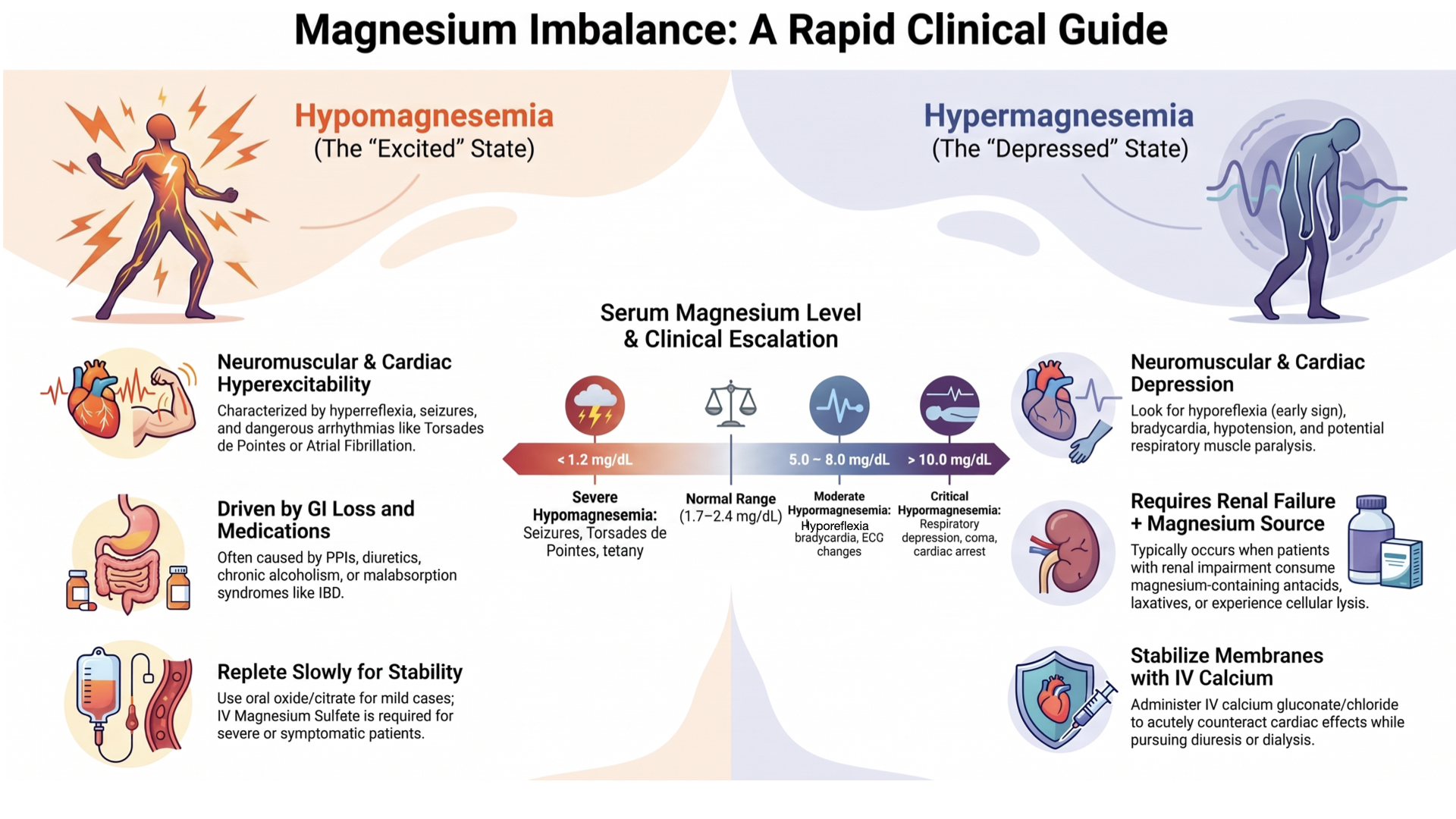
Normal Range: 1.7 – 2.4 mg/dL (0.7 – 1.0 mmol/L, 1.4 – 2.0 mEq/L) *
Conversion Factors:
- 1 mg/dL = 0.411 mmol/L = 0.833 mEq/L
- 1 mmol/L = 2.43 mg/dL = 2.0 mEq/L
- 1 mEq/L = 1.2 mg/dL = 0.5 mmol/L
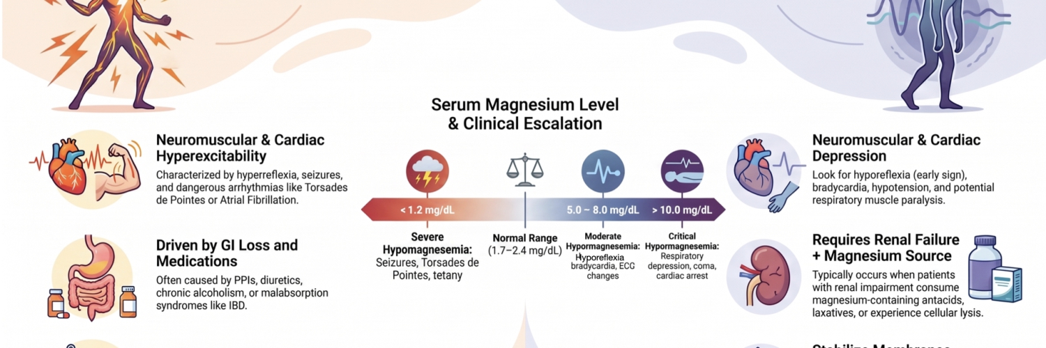
Hypomagnesemia
Definition & Epidemiology
- Defined as: Serum Mg < 1.7 mg/dL (0.7 mmol/L) *
- Prevalence: General population 3–10%; Type 2 diabetes 10–30%; Hospitalized 10–60%; ICU > 65% *
- Associated with: Low serum Mg2+ is an independent risk factor for long-term all-cause mortality in patients with non-ST-elevation myocardial infarction (NSTEMI) and may serve as a relatively inexpensive and fast biomarker for poor outcomes *.
Causes
- Medication-induced:
- Medications that cause GI loss of magnesium:
- Medications that cause intracellular shift of magnesium:
- Insulin.
- Beta-2 agonists (epinephrine, terbutaline, albuterol).
- Theophylline.
- Metformin.
- Alkali therapy *
- Medications that cause renal excretion of magnesium:
- Diuretics:
- Antibiotics:
- Aminoglycosides.
- Amphotericin B.
- Pentamidine.
- Foscarnet.
- Calcineurin inhibitors (tacrolimus > cyclosporine; hypomagnesemia may worsen adverse renal effects of the calcineurin inhibitor) *
- mTOR inhibitors (everolimus, sirolimus, temsirolimus).
- Platinum-based chemotherapy (cisplatin > carboplatin/oxaliplatin; may be long-lasting) *
- EGFR receptor blockers (e.g., cetuximab, erlotinib, matuzumab, panitumumab) — reduce TRPM6 activity
- Digoxin.
- Theophylline.
- Renal disease
- Chronic tubulointerstitial disease.
- Diuresis:
- Diuretics (discussed above).
- Post-ATN or post-obstructive (auto) diuresis.
- Osmotic diuresis (e.g., hyperglycemia).
- Renal tubular acidosis.
- Gastrointestinal disease
- Inadequate oral magnesium intake (common).
- Protein calorie malnutrition.
- Anorexia.
- Malabsorption:
- Inflammatory bowel disease.
- Short bowel syndrome.
- Vomiting, NG suction.
- Diarrhea.
- Pancreatitis (due to binding by free fatty acids; same mechanism that causes hypocalcemia).
- Medications
- Inadequate oral magnesium intake (common).
- Electrolyte abnormalities that cause hypomagnesemia
- Hypercalcemia.
- Hypophosphatemia.
- Specific clinical scenarios:
-
Hereditary causes: Mutations in TRPM6, TRPM7, CLDN16, CLDN19, NCC (Gitelman’s), EGF, EGFR, CNNM2 — account for ~80% of familial cases *
Clinical Manifestations
- Cardiovascular
- Prolonged QT, Torsades de pointes, atrial fibrillation, atrial flutter, multifocal atrial tachycardia, PVCs *
- Neuromuscular
- Tremors, fasciculations, cramps, hyperreflexia, tetany, carpopedal spasm, ⚡️seizures, delirium, psychosis, ataxia, nystagmus, dysarthria *
- Metabolic
- Insulin resistance, hypokalemia, hypocalcemia, metabolic alkalosis *
- Other:
- Patients with hypomagnesemia who are taking digitalis have an increased risk of developing Digitalis intoxication.
-
Low magnesium levels can increase myocardial sensitivity.
-
This increased sensitivity can lead to digitalis toxicity even when patients have a “normal” serum level of digitalis *.
-
- Patients with hypomagnesemia who are taking digitalis have an increased risk of developing Digitalis intoxication.
Associated Electrolyte Abnormalities
🟥 Hypokalemia
- Magnesium deficiency amplifies renal K⁺ loss via inhibition of Na⁺-K⁺-ATPase, ROMK channel opening, and NCC downregulation.
- Refractory hypokalemia corrects only after Mg repletion *.
🟥 Hypocalcemia
- Magnesium is essential for parathyroid hormone (PTH) function in two ways:
- PTH Release: The parathyroid glands need magnesium to secrete PTH. When magnesium is low, PTH release is suppressed (hypoparathyroidism).
- PTH Activity: Even if PTH is released, magnesium is required for it to work effectively on bone and kidneys to raise calcium.
- Result: Low magnesium → low PTH → low calcium (hypocalcemia).
- The Complication: Treating Hypomagnesemia Can Worsen Hypocalcemia. When you give IV MgSO₄👇
- Magnesium (Mg²⁺) and calcium (Ca²⁺) are both divalent cations.
- In the blood, Mg²⁺ can compete with Ca²⁺ for binding sites on proteins (like albumin).
- More importantly, Mg²⁺ can complex (bind) with free Ca²⁺ in the blood, forming an inactive complex.
- This transiently lowers the ionized (active) calcium level.
- In a patient who already has hypocalcemia, this temporary drop can be clinically significant, potentially causing worsening of symptoms like tetany, muscle spasms, or even arrhythmias.
- Clinical Implication
- Hypocalcemia that is refractory to calcium replacement alone is often due to underlying hypomagnesemia. Correcting the magnesium deficit is the missing piece *.
- Always check ionized calcium before and during magnesium repletion, especially in patients with known or suspected hypocalcemia.
- If calcium is critically low, consider giving IV calcium before or concurrently with magnesium to prevent a transient drop in ionized calcium.
- Monitor the patient closely for clinical signs of worsening hypocalcemia during magnesium infusion, including:
- Perioral or extremity paresthesias
- Muscle cramps or spasms
- Carpopedal spasm (positive Chvostek’s or Trousseau’s sign)
- Prolonged QT interval on ECG
- Seizures or altered mental status
- Understand that this effect is transient. The magnesium-calcium complexing is temporary. Once magnesium levels are restored, PTH function recovers, and calcium levels will improve over hours to days.
- Do not withhold magnesium out of fear of worsening hypocalcemia. The underlying cause (hypomagnesemia suppressing PTH) must be corrected. The key is to manage both electrolytes simultaneously with careful monitoring.
🟥 Thiamine deficiency
- It may arise via mechanisms similar to those implicated in magnesium deficiency (ie, inadequate intake, GI losses).
- Additionally, magnesium and thiamine are essential cofactors in critical metabolic pathways.
- Magnesium replacement should be considered when treating thiamine deficiency or conditions such as Wernicke encephalopathy or beriberi. Indeed, thiamine-refractory Wernicke encephalopathy that resolves with magnesium replacement has been described *.
Evaluation
-
Perform a complete metabolic panel.
-
Assess renal function, as well as potassium and calcium levels.
-
-
Perform an ECG.
-
Hypomagnesemia is commonly associated with a prolonged QT interval and may cause torsades de pointes.
-
-
Perform a head computed tomography (CT) scan.
-
Consider assessing the patient for alternate causes of altered mental status or seizures.
-
Management
General principles
- Mg is generally safe; caution in myasthenia gravis and GFR < 30 ml/min *.
- ⚠️Patients with myasthenia gravis may be at increased risk of muscle weakness.
 Renal failure (e.g. GFR < 30 ml/min) may cause magnesium accumulation. These patients may be treated with a normal “loading” dose of magnesium up-front, but care is needed with repeated dosing.
- Magnesium repletion can be difficult:
- Oral magnesium is poorly absorbed and causes diarrhea.
- IV magnesium boluses cause a transient elevation in the serum magnesium level, which causes magnesium secretion by the kidneys. Most of the administered magnesium may be excreted in the urine.
- Most of the body’s magnesium is intracellular. The goal is to get magnesium into the cells, but cellular uptake occurs slowly.
- Adjuvant therapies (refractory)
Oral Mg Repletion
◾️Indication: Oral magnesium may be used to treat hypomagnesemia if all of the following criteria are met:
- Hypomagnesemia is mild (e.g., ~1.6-2 mg/dL), or asymptomatic.
- The patient is taking oral medications
- There is no interaction with other medications (e.g., tetracyclines and calcium channel blockers).
◾️Side effect: Diarrhea
💊 Formulation & disong
- Magnesium oxide, Magnesium citrate, Magnesium gluconate, Magnesium Supplements (Mg-Tab SR)
- Organic salts (citrate, glycinate, gluconate) have better bioavailability (25–40%) than inorganic salts (oxide ~20–30%) *
- Dosage and consideration: See following table
IV Mg Repletion
Contraindication
- ⛔️Heart block.
- ⛔️Severe hypocalcemia.
- ⚠️Myasthenia gravis:
- Relative contraindication. Avoid if possible (e.g., use oral magnesium).
- For severe hypomagnesemia, intravenous magnesium can be utilized, but it should be given slowly and with extreme caution.
- ⚠️Renal failure:
- Magnesium may still be given, but the dose needs to be adjusted and monitored more carefully.
- GFR < 30 ml/min is a contraindication to a continuous magnesium infusion. If indicated, Mg should be given as intermittent boluses based on serum level.
Indications
- Hypomagnesemia
- Acute asthma
- Torsade de pointes
- AF with RVR *
- Preeclampsia/eclampsia
Administration
📈 Rate of infusion:
- Traditionally, magnesium sulfate is given slowly due to the side effects of rapid magnesium infusion (ie, hypotension, respiratory depression); however, in patients with severe clinical manifestations (e.g., hemodynamically unstable patients with arrhythmias, Tdps, or symptomatic hypokalemia), magnesium sulfate should be administered rapidly to prevent clinical decompensation *.
💉Dosing For Various Clinical Conditions
- Asymptomatic hypomagnesemia with Mg ~ 1.2-1.8 mg/dL
- Intermittent administration of 2-4 grams of magnesium sulfate IV.
- Recommended dosing for patients with GFR >50 ml/min:
- Mild hypomagnesemia (1.6-1.8 mg/dL): weight/16 = grams of IV magnesium sulfate over 24 hrs.
- Moderate hypomagnesemia (1.2-1.5 mg/dL): weight/8 = grams of IV magnesium sulfate over 24 hrs.
- Transitioning to scheduled oral magnesium may help prevent recurrence.
- Asymptomatic hypomagnesemia with Mg < 1.2 mg/dL
- Severe hypomagnesemia reflects a low total body Mg content.
- Possible options for Mg repletion:
- Multiple scheduled doses of IV magnesium (e.g., 2-4 grams IV magnesium sulfate q6hr-q8hr).
- Continuous infusion of IV magnesium.
- The recommended dose for patients with a GFR > 50 ml/min = weight/4 as the grams of IV magnesium over 24 hours
- Follow extended electrolytes (electrolytes plus Ca/Mg/Phos).
- 🚨Aggressive IV MgSO4 therapy (usually continuous infusion)
- Indications: Torsades de pointes, AF with RVR *, preeclampsia/eclampsia (below), severe hypomagnesemia with symptoms (⚡️seizures)
- Loading Dose: 4 g total — 2 g IV over 5–15 min, then 2 g IV over 30–60 min *, *
- Maintenance if (GFR > 30):1 g/h infusion for 24 hours. 👉If GFR < 30: Follow levels, re-dose based on level — “No Continuous Infusion“.
- Monitor electrolytes and serum Mg q6 hours for 24 hours
- Target serum Mg 3.6–4.9 mg/dL.
- If Mg 5–7 → Reduce infusion by 50%
- If > 7 → Stop.
- Monitor: Weakness/somnolence → check Mg; bradycardia/dyspnea → stop infusion and check Mg
- Preeclampsia/eclampsia
- Loading dose: IV MgSO4 4–6 g loading * , *
- Maintenance dose:
- If 1.1 < serum creatinine < 2.5 mg/dL: Continuous infusion of 1g/h *.
- ⚠️If serum creatinine ≥2.5 mg/dL or severe oliguria: Do not give a maintenance infusion.
- Follow electrolytes & magnesium levels q4-6hr. Give magnesium bolus based on serum level
- Monitor for features of magnesium toxicity *
- Duration of therapy: Treatment is often continued until ~24 hours after delivery, when the patient shows clinical resolution of preeclampsia.
- Acute asthma
- IV MgSO4 2 g IV over 20 min (guidelines recommend for severe exacerbations) *
Hypermagnesemia
Definition & Epidemiology
-
Defined as: Serum Mg > 2.4 mg/dL (> 1.0 mmol/L) *
-
Rare: Occurs almost exclusively in patients with renal impairment receiving magnesium-containing products *
- Note ↑Magnesium is closely associated with ↑K and ↓Ca
Causes
- Exogenous magnesium: Mg infusions (preeclampsia), Mg-containing laxatives, antacids, enemas *
- Hypermagnesemia has also been described in patients with obstipation and fecal impaction, both with and without the administration of magnesium-containing laxatives.
- Endogenous release: Rhabdomyolysis, hemolysis, tumor lysis syndrome, crush injury, severe burns
- Pseudo-hypermagnesemia: Hemolyzed blood sample → repeat lab
📍Key point: Persistent hypermagnesemia requires renal failure plus an ongoing Mg source
Clinical Manifestations
- 4–5 mg/dL: Lethargy, nausea, headache, flushing
- 5–8 mg/dL: Hyporeflexia, hypotension, bradycardia, ECG changes (wide QRS, peaked T-waves)
- 8–10 mg/dL: Muscle weakness, drowsiness, AV block, progressive respiratory depression
- 10–15 mg/dL: Absent reflexes, respiratory depression, coma
- > 15 mg/dL: Cardiac arrest, complete heart block *
Physical Examination
- Hyporeflexia: Early sign; presence of reflexes argues against severe hypermagnesemia (but nonspecific)
- Note: Decreased tendon reflexes are generally seen with serum magnesium levels >10 mg/dL (>4 mmol/L), although they can be seen at serum magnesium levels as low as 5 mg/dL (2 mmol/L).
- Bradycardia, hypotension *
ECG Findings
- ECG (as for hyperkalaemia)
- Increased PR and QTc
- Wide QRS
- Peaked T waves and flattened P waves
- Complete AV block and asystole, heart block *
Evaluation
- Complete electrolytes (Ca/Mg/Phos)
- LDH, CK, or uric acid (rhabdomyolysis, hemolysis, tumor lysis)
- Rule out hemolyzed sample
Management
Step 1: Membrane Stabilization with IV Calcium
- For severe, symptomatic hypermagnesemia (cardiac dysfunction, respiratory muscle paralysis, or significant neurologic symptoms)
- Calcium gluconate 2 gr IV over 5–10 minutes (or 1 gram calcium chloride)
- May need to repeat
- Mechanism: Acutely counteracts cardiac and neuromuscular effects of hypermagnesemia
Step 2: Volume Resuscitation
- If patient is hypovolemic, resuscitate with magnesium-free IV fluids
- Use: Lactated Ringer’s or normal saline
- Note: Dilutional effect could theoretically lower serum Mg, but evidence is lacking
Step 3: Diuresis (Loop + Thiazide)
- For patients with preserved renal function
- Strategy: Loop diuretic (e.g., furosemide) + thiazide diuretic (IV chlorothiazide or oral metolazone)
- Rationale: Dual-site nephron blockade — loop diuretic alone may not be sufficient as distal tubule can still reclaim Mg
- Replete urine losses with Mg-free IV fluids to maintain even fluid balance
- Monitor electrolytes carefully (Ca/Mg/Phos, ionized calcium)
- ⚠️ No high-level evidence supports this strategy
Step 4: Specific Therapy for Underlying Cause
- Treat the underlying condition causing cellular lysis:
- Rhabdomyolysis — aggressive hydration, monitor renal function
- Tumor lysis syndrome — rasburicase, hydration, other supportive measures
- Hemolysis — manage underlying cause
- Crush injury, severe burns — supportive care
Step 5: Dialysis
- Definitive treatment for hypermagnesemia in patients with anuric renal failure
- Highly effective at removing magnesium
- Indication: Severe hypermagnesemia with renal failure where other measures are insufficient *.
RECAP
-
Hypomagnesemia common, hypermagnesemia rare. Suspect hypomagnesemia in ICU patients, diabetics, alcohol use disorder, and patients on PPIs, diuretics, cisplatin, or EGFR inhibitors *.
-
Serum Mg does not reflect total body stores. < 1% of total body Mg is in plasma. “Normal” serum level does not rule out deficiency *.
-
Refractory hypokalemia and hypocalcemia require Mg repletion. Neither will correct until Mg deficiency is addressed *.
-
IV Mg boluses are rapidly excreted. Continuous infusion is more effective for severe depletion.
-
Hypermagnesemia is a renal failure story. Persistent hypermagnesemia always requires renal impairment plus an exogenous or endogenous Mg source *.
-
Magnesium is a therapeutic agent of choice for:
- AF with RVR
- Torsades de pointes
- Preeclampsia/eclampsia
- Acute asthma exacerbations
References
1. PMID: 38838313. Touyz RM, de Baaij JHF, Hoenderop JGJ. Magnesium Disorders. N Engl J Med. 2024 Jun 6;390(21):1998-2009. doi: 10.1056/NEJMra1510603.
2. PMID: 38372687. Adomako EA, Yu ASL. Magnesium Disorders: Core Curriculum 2024. Am J Kidney Dis. 2024 Jun;83(6):803-815. doi: 10.1053/j.ajkd.2023.10.017. Epub 2024 Feb 17.
3. PMID: 36872194. Rosner MH, Ha N, Palmer BF, Perazella MA. Acquired Disorders of Hypomagnesemia. Mayo Clin Proc. 2023 Apr;98(4):581-596. doi: 10.1016/j.mayocp.2022.12.002. Epub 2023 Mar 3.
4. PMID: 30220246. Van Laecke S. Hypomagnesemia and hypermagnesemia. Acta Clin Belg. 2019 Feb;74(1):41-47. doi: 10.1080/17843286.2018.1516173. Epub 2018 Sep 17.
5. PMID: 24402002. Ayuk J, Gittoes NJ. Contemporary view of the clinical relevance of magnesium homeostasis. Ann Clin Biochem. 2014 Mar;51(Pt 2):179-88. doi: 10.1177/0004563213517628. Epub 2014 Jan 8.

Add comment